Zhu Lei, Liu Ya-Jun, Shen Hong, Gu Pei-Qing, Zhang Lu
Jiangsu Province Hospital of Traditional Chinese Medicine (TCM), Affiliated Hospital of Nanjing University of Traditional Chinese Medicine (TCM), Nanjing, Jiangsu, China (mainland).
Jiangsu Province Hospital of Traditional Chinese Medicine (TCM), Affiliated Hospital of Nanjing University of TCM, Nanjing, Jiangsu, China (mainland).
Med Sci Monit. 2017 Jul 1;23:3209-3216. doi: 10.12659/msm.902441.
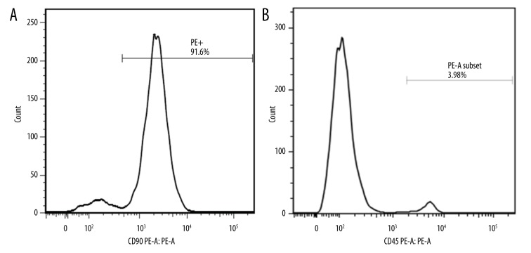
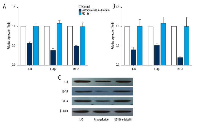
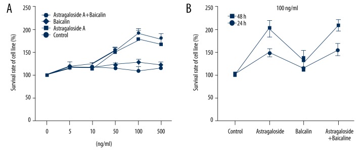
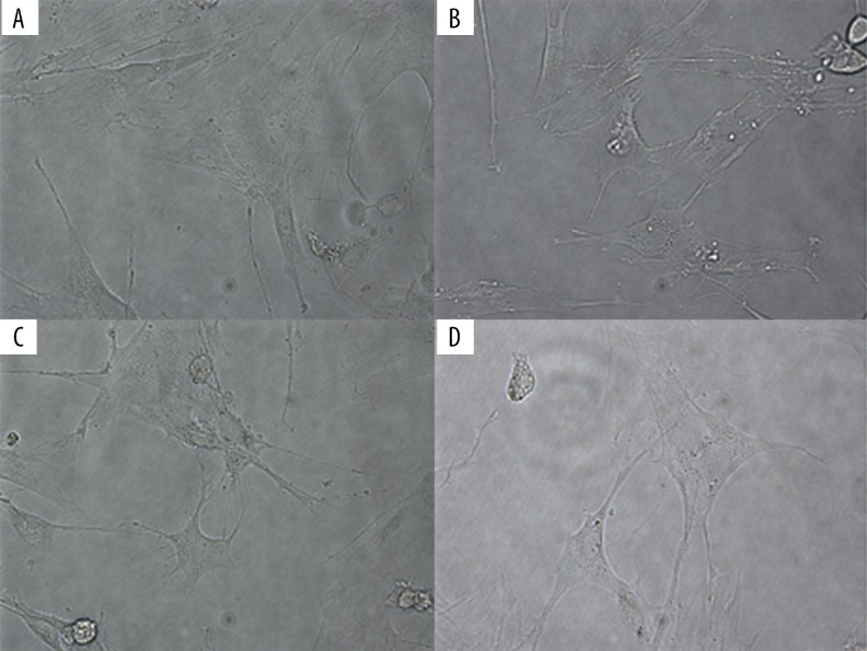
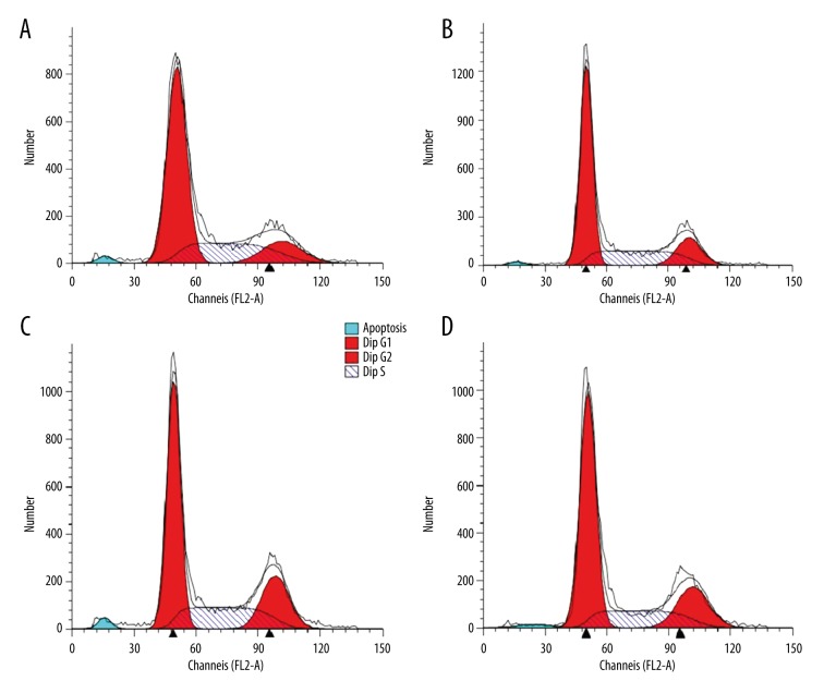
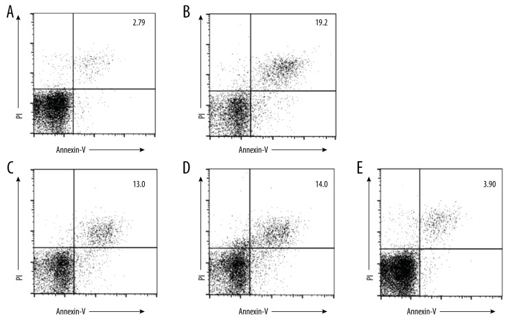
BACKGROUND Mesenchymal stem cells (MSCs) have emerged as an attractive alternative to modulating immune response after transplantation. Recent studies have shown that systemically administered MSCs enter the inflamed intestine. In the present study, we propose a strategy to improve the efficacy of MSC-based cellular therapy for inflammation using Astragaloside and Baicalein to enhance cell survival, inhibit apoptosis, and modulate inflammatory response in vitro. MATERIAL AND METHODS MSCs were induced with lipopolysaccharide (LPS) as an inflammatory model before being treated for 48 h with Astragaloside, Baicalein, and the combination of both. MSCs proliferation was determined using the MTT method. The cell cycle situation was monitored using flow cytometry, and the apoptosis ability of MSCs was detected with Annexin-V flow cytometry. The levels of cytokine IL-1β, IL-8, and TNF-α, and their relations with the ERK pathway were measured using ELISA, RT-PCR, and Western blot. RESULTS Compared to the control groups (containing no drug), each drug-treated group showed the ability to promote epithelial differentiation and cell growth and to inhibit apoptosis. The combination group had reduced levels of IL-1β, IL-8, and TNF-α in LPS-induced MSCs, much more than in the other 2 groups. Compared with the other groups, the combination of Astragaloside and Baicalin more efficiently reduced IL-1β, IL-8, and TNF-α levels in the LPS-induced MSCs model, and ERK inhibitor was capable of recovering the inflammatory effect. CONCLUSIONS The results demonstrated that Astragaloside and Baicalin can promote epithelial differentiation and proliferation, inhibit apoptosis, and reduce inflammatory effects.
背景 间充质干细胞(MSCs)已成为移植后调节免疫反应的一种有吸引力的替代方法。最近的研究表明,全身给药的间充质干细胞可进入发炎的肠道。在本研究中,我们提出了一种策略,使用黄芪甲苷和黄芩苷来提高基于间充质干细胞的细胞疗法对炎症的疗效,以增强细胞存活、抑制细胞凋亡并在体外调节炎症反应。材料与方法 以脂多糖(LPS)诱导间充质干细胞作为炎症模型,然后用黄芪甲苷、黄芩苷及其组合处理48小时。使用MTT法测定间充质干细胞的增殖。使用流式细胞术监测细胞周期情况,并用膜联蛋白-V流式细胞术检测间充质干细胞的凋亡能力。使用酶联免疫吸附测定(ELISA)、逆转录-聚合酶链反应(RT-PCR)和蛋白质免疫印迹法测量细胞因子IL-1β、IL-8和TNF-α的水平及其与细胞外调节蛋白激酶(ERK)途径的关系。结果 与对照组(不含药物)相比,各药物处理组均显示出促进上皮分化和细胞生长以及抑制细胞凋亡的能力。联合组在LPS诱导的间充质干细胞中IL-1β、IL-8和TNF-α的水平降低,比其他两组降低得多。与其他组相比,黄芪甲苷和黄芩苷的组合在LPS诱导的间充质干细胞模型中更有效地降低了IL-1β、IL-8和TNF-α的水平,并且ERK抑制剂能够恢复炎症效应。结论 结果表明,黄芪甲苷和黄芩苷可促进上皮分化和增殖,抑制细胞凋亡,并减轻炎症效应。