School of Biosciences, University of Exeter, Exeter, UK.
Department of Biology, University of Utrecht, Utrecht, The Netherlands.
Cell Microbiol. 2017 Nov;19(11). doi: 10.1111/cmi.12764. Epub 2017 Aug 9.
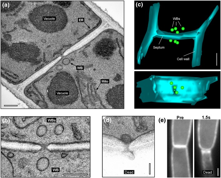
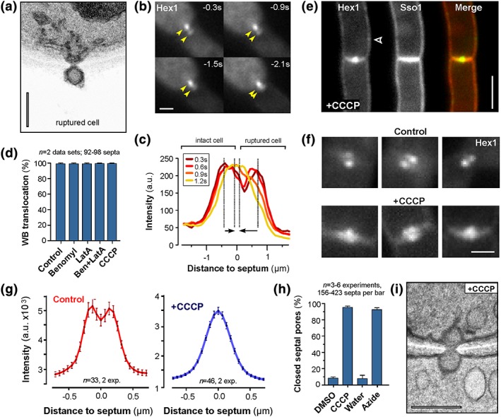
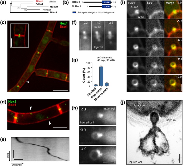
Septa of filamentous ascomycetes are perforated by septal pores that allow communication between individual hyphal compartments. Upon injury, septal pores are plugged rapidly by Woronin bodies (WBs), thereby preventing extensive cytoplasmic bleeding. The mechanism by which WBs translocate into the pore is not known, but it has been suggested that wound-induced cytoplasmic bleeding "flushes" WBs into the septal opening. Alternatively, contraction of septum-associated tethering proteins may pull WBs into the septal pore. Here, we investigate WB dynamics in the wheat pathogen Zymoseptoria tritici. Ultrastructural studies showed that 3.4 ± 0.2 WBs reside on each side of a septum and that single WBs of 128.5 ± 3.6 nm in diameter seal the septal pore (41 ± 1.5 nm). Live cell imaging of green fluorescent ZtHex1, a major protein in WBs, and the integral plasma membrane protein ZtSso1 confirms WB translocation into the septal pore. This was associated with the occasional formation of a plasma membrane "balloon," extruding into the dead cell, suggesting that the plasma membrane rapidly seals the wounded septal pore wound. Minor amounts of fluorescent ZtHex1-enhanced green fluorescent protein (eGFP) appeared associated with the "ballooning" plasma membrane, indicating that cytoplasmic ZtHex1-eGFP is recruited to the extending plasma membrane. Surprisingly, in ~15% of all cases, WBs moved from the ruptured cell into the septal pore. This translocation against the cytoplasmic flow suggests that an active mechanism drives WB plugging. Indeed, treatment of unwounded and intact cells with the respiration inhibitor carbonyl cyanide m-chlorophenyl hydrazone induced WB translocation into the pores. Moreover, carbonyl cyanide m-chlorophenyl hydrazone treatment recruited cytoplasmic ZtHex1-eGFP to the lateral plasma membrane of the cells. Thus, keeping the WBs out of the septal pores, in Z. tritici, is an ATP-dependent process.
丝状子囊菌的隔膜被隔膜孔穿孔,允许单个菌丝隔室之间进行通信。受伤后,隔膜孔很快被Woronin 体(WB)堵塞,从而防止细胞质广泛出血。WB 转移到孔中的机制尚不清楚,但有人提出,伤口诱导的细胞质出血“冲洗”WB 进入隔膜开口。或者,隔膜相关系绳蛋白的收缩可能会将 WB 拉入隔膜孔。在这里,我们研究了小麦病原体小麦纹枯病菌中的 WB 动态。超微结构研究表明,每个隔膜的两侧有 3.4±0.2 个 WB,直径为 128.5±3.6nm 的单个 WB 密封隔膜孔(41±1.5nm)。绿色荧光 ZtHex1(WB 中的主要蛋白质)和完整质膜蛋白 ZtSso1 的活细胞成像证实了 WB 向隔膜孔的易位。这与质膜“气球”偶尔形成有关,质膜向外突出到死细胞中,表明质膜迅速密封受伤的隔膜孔伤口。少量荧光 ZtHex1 增强型绿色荧光蛋白(eGFP)似乎与“气球”质膜相关,表明细胞质 ZtHex1-eGFP 被募集到延伸的质膜上。令人惊讶的是,在所有情况下的约 15%中,WB 从破裂的细胞移动到隔膜孔中。这种与细胞质流相反的易位表明一种活跃的机制驱动 WB 堵塞。事实上,用呼吸抑制剂羰基氰化物 m-氯苯腙处理未受伤和完整的细胞会诱导 WB 向孔中易位。此外,羰基氰化物 m-氯苯腙处理会将细胞质 ZtHex1-eGFP 募集到细胞的侧质膜上。因此,在小麦纹枯病菌中,保持 WB 不在隔膜孔中是一个依赖于 ATP 的过程。