Suppr超能文献

全身狄克逊磁共振成像在多发性骨髓瘤中的诊断效用:一项多阅片者研究

Diagnostic utility of whole body Dixon MRI in multiple myeloma: A multi-reader study.

作者信息

Bray Timothy J P, Singh Saurabh, Latifoltojar Arash, Rajesparan Kannan, Rahman Farzana, Narayanan Priya, Naaseri Sahar, Lopes Andre, Bainbridge Alan, Punwani Shonit, Hall-Craggs Margaret A

机构信息

Centre for Medical Imaging, University College London, London, United Kingdom.

Cancer Research UK and UCL Clinical Trials Centre, London, United Kingdom.

出版信息

PLoS One. 2017 Jul 3;12(7):e0180562. doi: 10.1371/journal.pone.0180562. eCollection 2017.

Abstract

OBJECTIVE

To determine which of four Dixon image types [in-phase (IP), out-of-phase (OP), fat only (FO) and water-only (WO)] is most sensitive for detecting multiple myeloma (MM) focal lesions on whole body MRI (WB-MRI) images.

METHODS

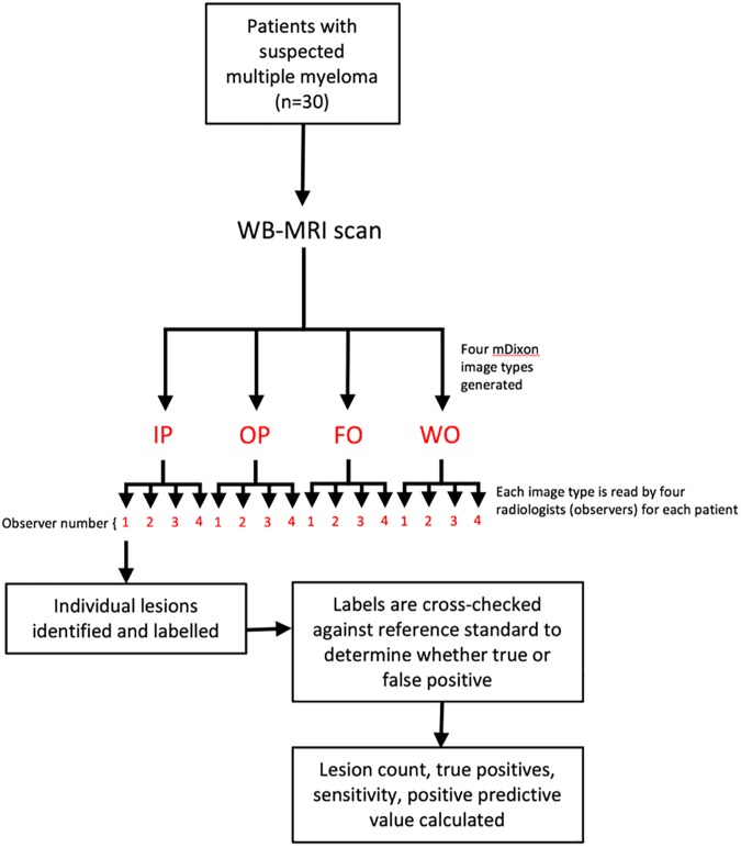
Thirty patients with clinically-suspected MM underwent WB-MRI at 3 Tesla. Unenhanced IP, OP, FO and WO Dixon images were generated and read by four radiologists. On each image type, each radiologist identified and labelled all visible myeloma lesions in the bony pelvis. Each identified lesion was compared with a reference standard consisting of pre- and post-contrast Dixon and diffusion weighted imaging (read by a further consultant radiologist) to determine whether the lesion was truly positive. Lesion count, true positives, sensitivity, and positive predictive value were compared across the four Dixon image types.

RESULTS

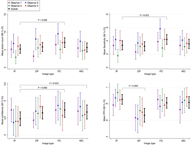
Lesion count, true positives, sensitivity and confidence scores were all significantly higher on FO images than on IP images (p>0.05).

DISCUSSION

FO images are more sensitive than other Dixon image types for MM focal lesions, and should be preferentially read by radiologists to improve diagnostic accuracy and reporting efficiency.

摘要

目的

确定四种 Dixon 图像类型(同相位(IP)、反相位(OP)、仅脂肪(FO)和仅水(WO))中哪一种对全身 MRI(WB-MRI)图像上多发性骨髓瘤(MM)局灶性病变的检测最为敏感。

方法

30 例临床疑似 MM 的患者在 3 特斯拉磁场下接受 WB-MRI 检查。生成未增强的 IP、OP、FO 和 WO Dixon 图像,并由四名放射科医生进行解读。在每种图像类型上,每位放射科医生在骨盆骨中识别并标记所有可见的骨髓瘤病变。将每个识别出的病变与由对比剂前后 Dixon 图像和扩散加权成像组成的参考标准(由另一位放射科顾问医生解读)进行比较,以确定该病变是否为真正的阳性病变。比较四种 Dixon 图像类型的病变数量、真阳性、敏感性和阳性预测值。

结果

FO 图像上的病变数量、真阳性、敏感性和可信度评分均显著高于 IP 图像(p>0.05)。

讨论

FO 图像对 MM 局灶性病变比其他 Dixon 图像类型更敏感,放射科医生应优先解读 FO 图像以提高诊断准确性和报告效率。

https://cdn.ncbi.nlm.nih.gov/pmc/blobs/e516/5495520/0d18fd769755/pone.0180562.g001.jpg

相似文献

1
Diagnostic utility of whole body Dixon MRI in multiple myeloma: A multi-reader study.
PLoS One. 2017 Jul 3;12(7):e0180562. doi: 10.1371/journal.pone.0180562. eCollection 2017.
2
Whole body MRI in multiple myeloma: Optimising image acquisition and read times.
PLoS One. 2020 Jan 30;15(1):e0228424. doi: 10.1371/journal.pone.0228424. eCollection 2020.
4
Improved contrast for myeloma focal lesions with T2-weighted Dixon images compared to T1-weighted images.
Diagn Interv Imaging. 2019 Sep;100(9):513-519. doi: 10.1016/j.diii.2019.05.001. Epub 2019 May 23.
5
Optimization of whole-body 2-[F]FDG-PET/MRI imaging protocol for the initial staging of patients with myeloma.
Eur Radiol. 2022 May;32(5):3085-3096. doi: 10.1007/s00330-021-08388-6. Epub 2021 Nov 29.
8
Conspicuity of bone metastases on fast Dixon-based multisequence whole-body MRI: clinical utility per sequence.
Magn Reson Imaging. 2013 Jun;31(5):669-75. doi: 10.1016/j.mri.2012.10.017. Epub 2013 Jan 3.

引用本文的文献

1
Recent advances in imaging and artificial intelligence (AI) for quantitative assessment of multiple myeloma.
Am J Nucl Med Mol Imaging. 2024 Aug 25;14(4):208-229. doi: 10.62347/NLLV9295. eCollection 2024.
2
Review of whole-body magnetic resonance imaging in multiple myeloma.
Jpn J Radiol. 2024 Dec;42(12):1381-1391. doi: 10.1007/s11604-024-01635-y. Epub 2024 Aug 1.
3
New diagnostic strategy for multiple myeloma: A review.
Medicine (Baltimore). 2023 Dec 29;102(52):e36660. doi: 10.1097/MD.0000000000036660.
4
Whole-Body MRI in Musculoskeletal Oncology: A Comprehensive Review with Recommendations.
Radiol Imaging Cancer. 2023 May;5(3):e220107. doi: 10.1148/rycan.220107.
6
Advanced Imaging in Multiple Myeloma: New Frontiers for MRI.
Diagnostics (Basel). 2022 Sep 9;12(9):2182. doi: 10.3390/diagnostics12092182.
7
Imaging of treatment response and minimal residual disease in multiple myeloma: state of the art WB-MRI and PET/CT.
Skeletal Radiol. 2022 Jan;51(1):59-80. doi: 10.1007/s00256-021-03841-5. Epub 2021 Aug 7.
9
Diagnostic Value of Whole-Body MRI Short Protocols in Bone Lesion Detection in Multiple Myeloma Patients.
Diagnostics (Basel). 2021 Jun 8;11(6):1053. doi: 10.3390/diagnostics11061053.

本文引用的文献

1
Whole-Body MR Imaging: Musculoskeletal Applications.
Radiology. 2016 May;279(2):345-65. doi: 10.1148/radiol.2016142084.
2
Treatment of multiple myeloma with high-risk cytogenetics: a consensus of the International Myeloma Working Group.
Blood. 2016 Jun 16;127(24):2955-62. doi: 10.1182/blood-2016-01-631200. Epub 2016 Mar 21.
5
International Myeloma Working Group updated criteria for the diagnosis of multiple myeloma.
Lancet Oncol. 2014 Nov;15(12):e538-48. doi: 10.1016/S1470-2045(14)70442-5. Epub 2014 Oct 26.
7
Whole-body diffusion-weighted MR imaging for assessment of treatment response in myeloma.
Radiology. 2014 Jun;271(3):785-94. doi: 10.1148/radiol.13131529. Epub 2014 Jan 21.
8
Prognostic significance of whole-body MRI in patients with monoclonal gammopathy of undetermined significance.
Leukemia. 2014 Jan;28(1):174-8. doi: 10.1038/leu.2013.244. Epub 2013 Aug 20.
10
Conspicuity of bone metastases on fast Dixon-based multisequence whole-body MRI: clinical utility per sequence.
Magn Reson Imaging. 2013 Jun;31(5):669-75. doi: 10.1016/j.mri.2012.10.017. Epub 2013 Jan 3.

文献AI研究员

20分钟写一篇综述,助力文献阅读效率提升50倍。

立即体验

用中文搜PubMed

大模型驱动的PubMed中文搜索引擎

马上搜索

文档翻译

学术文献翻译模型,支持多种主流文档格式。

立即体验