Hojo Hidehiro, Dohmae Takeshi, Hotta Kenji, Kohno Ryosuke, Motegi Atsushi, Yagishita Atsushi, Makinoshima Hideki, Tsuchihara Katsuya, Akimoto Tetsuo
Division of Radiation Oncology and Particle Therapy, National Cancer Center Hospital East, 6-5-1, Kashiwanoha, Kashiwa, Chiba, 277-8577, Japan.
High Energy Accelerator Research Organization, 1-1 Oho, Tsukuba, Ibaraki, 305-0801, Japan.
Radiat Oncol. 2017 Jul 3;12(1):111. doi: 10.1186/s13014-017-0849-1.
Cellular responses to proton beam irradiation are not yet clearly understood, especially differences in the relative biological effectiveness (RBE) of high-energy proton beams depending on the position on the Spread-Out Bragg Peak (SOBP). Towards this end, we investigated the differences in the biological effect of a high-energy proton beam on the target cells placed at different positions on the SOBP, using two human esophageal cancer cell lines with differing radiosensitivities.
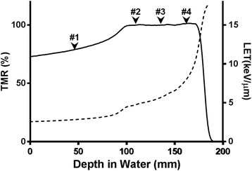
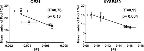
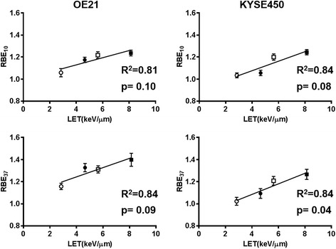
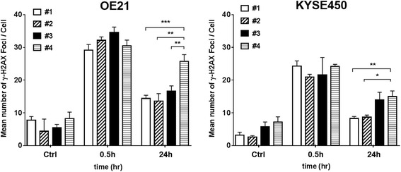
Two human esophageal cancer cell lines (OE21, KYSE450) with different radiosensitivities were irradiated with a 235-MeV proton beam at 4 different positions on the SOBP (position #1: At entry; position #2: At the proximal end of the SOBP; position #3: Center of the SOBP; position #4: At the distal end of the SOBP), and the cell survivals were assessed by the clonogenic assay. The RBE for each position of the target cell lines on the SOBP was determined based on the results of the cell survival assay conducted after photon beam irradiation. In addition, the number of DNA double-strand breaks was estimated by quantitating the number of phospho-histone H2AX (γH2AX) foci formed in the nuclei by immunofluorescence analysis.
In regard to differences in the RBE of a proton beam according to the position on the SOBP, the RBE value tended to increase as the position on the SOBP moved distally. Comparison of the residual number of γH2AX foci at the end 24 h after the irradiation revealed, for both cell lines, a higher number of foci in the cells irradiated at the distal end of the SOPB than in those irradiated at the proximal end or center of the SOBP.
The results of this study demonstrate that the RBE of a high-energy proton beam and the cellular responses, including the DNA damage repair processes, to high-energy proton beam irradiation, differ according to the position on the SOBP, irrespective of the radiosensitivity levels of the cell lines.
细胞对质子束照射的反应尚未完全明确,尤其是高能质子束的相对生物效应(RBE)因在扩展布拉格峰(SOBP)上的位置不同而存在差异。为此,我们使用两种放射敏感性不同的人食管癌细胞系,研究了高能质子束对置于SOBP不同位置的靶细胞的生物学效应差异。
用235 MeV的质子束在SOBP的4个不同位置(位置#1:入射处;位置#2:SOBP近端;位置#3:SOBP中心;位置#4:SOBP远端)照射两种放射敏感性不同的人食管癌细胞系(OE21、KYSE450),并通过克隆形成试验评估细胞存活率。根据光子束照射后进行的细胞存活试验结果,确定靶细胞系在SOBP各位置的RBE。此外,通过免疫荧光分析定量细胞核中形成的磷酸化组蛋白H2AX(γH2AX)灶的数量,估计DNA双链断裂的数量。
关于质子束RBE根据SOBP上位置的差异,RBE值倾向于随着SOBP上的位置向远端移动而增加。照射后24小时结束时γH2AX灶残留数量的比较显示,对于两种细胞系,在SOBP远端照射的细胞中形成的灶数量均高于在SOBP近端或中心照射的细胞。
本研究结果表明,高能质子束的RBE以及细胞对高能质子束照射的反应,包括DNA损伤修复过程,根据SOBP上的位置而有所不同,与细胞系的放射敏感性水平无关。