Department of Psychiatry, Yale University School of Medicine, New Haven, Connecticut; Department of Psychiatry and Behavioral Neuroscience, University of Cincinnati College of Medicine, Cincinnati, Ohio.
Department of Psychiatry, Yale University School of Medicine, New Haven, Connecticut.
Biol Psychiatry. 2018 Jan 1;83(1):38-49. doi: 10.1016/j.biopsych.2017.05.026. Epub 2017 Jun 12.
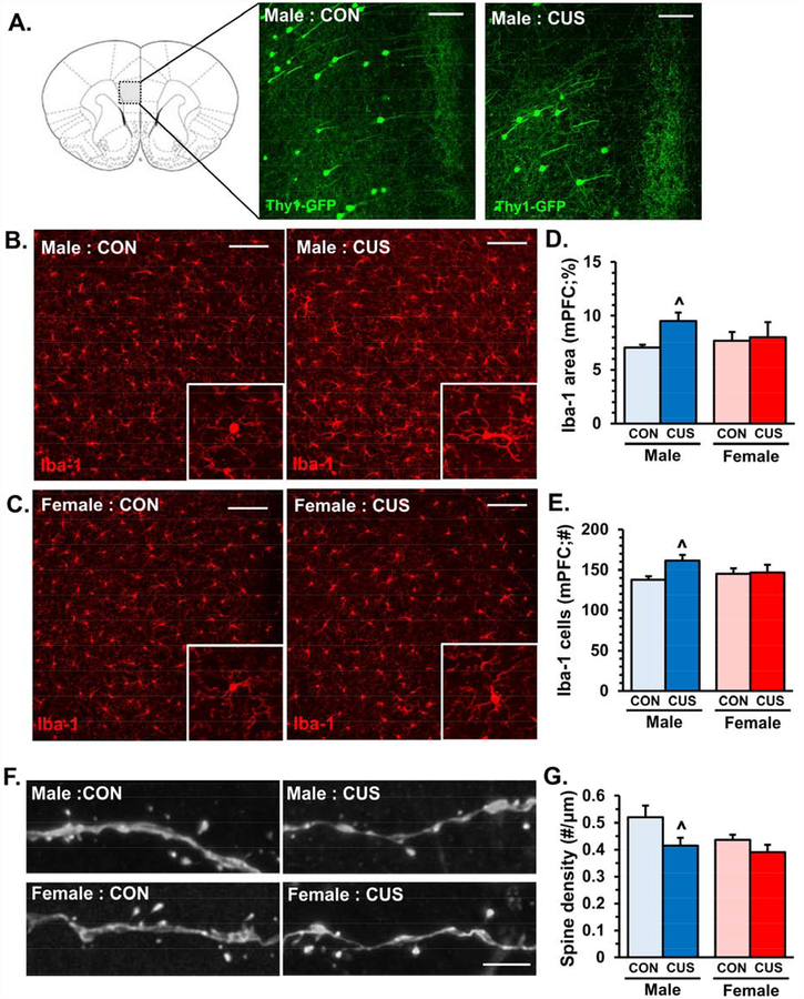
Chronic stress exposure causes neuronal atrophy and synaptic deficits in the medial prefrontal cortex (PFC), contributing to development of anxiety- and depressive-like behaviors. Concomitantly, microglia in the PFC undergo morphological and functional changes following stress exposure, suggesting that microglia contribute to synaptic deficits underlying behavioral consequences.
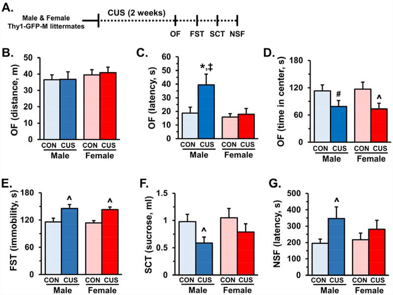
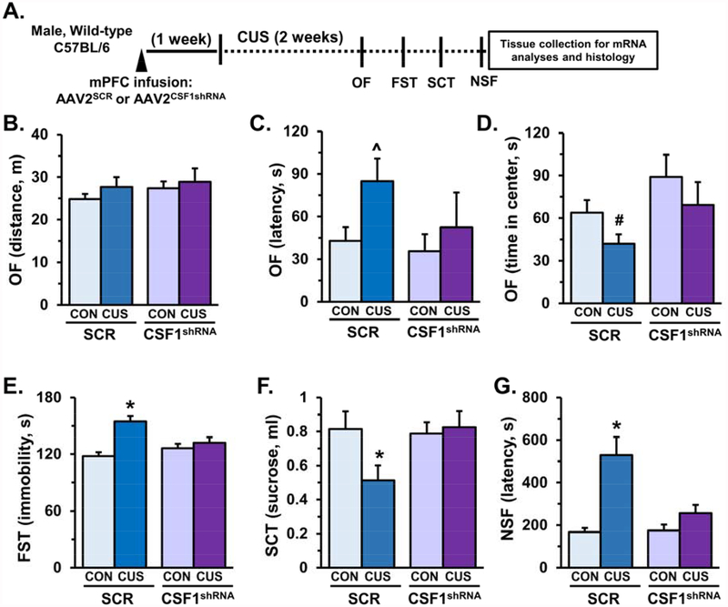
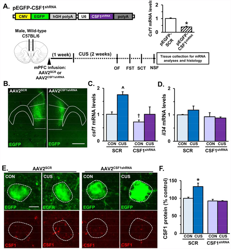
Male and female mice were exposed to chronic unpredictable stress (CUS) to examine the role of neuron-microglia interactions in the medial PFC during development of anxiety- and depressive-like behaviors. Thy1-GFP-M mice were used to assess microglia-mediated neuronal remodeling and dendritic spine density in the medial PFC. Viral-mediated knockdown of neuronal colony stimulating factor 1 (CSF1) was used to modulate microglia function and behavioral consequences after CUS.
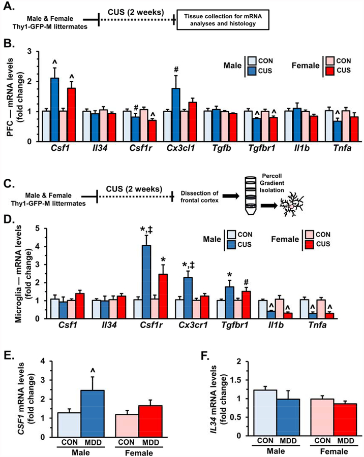
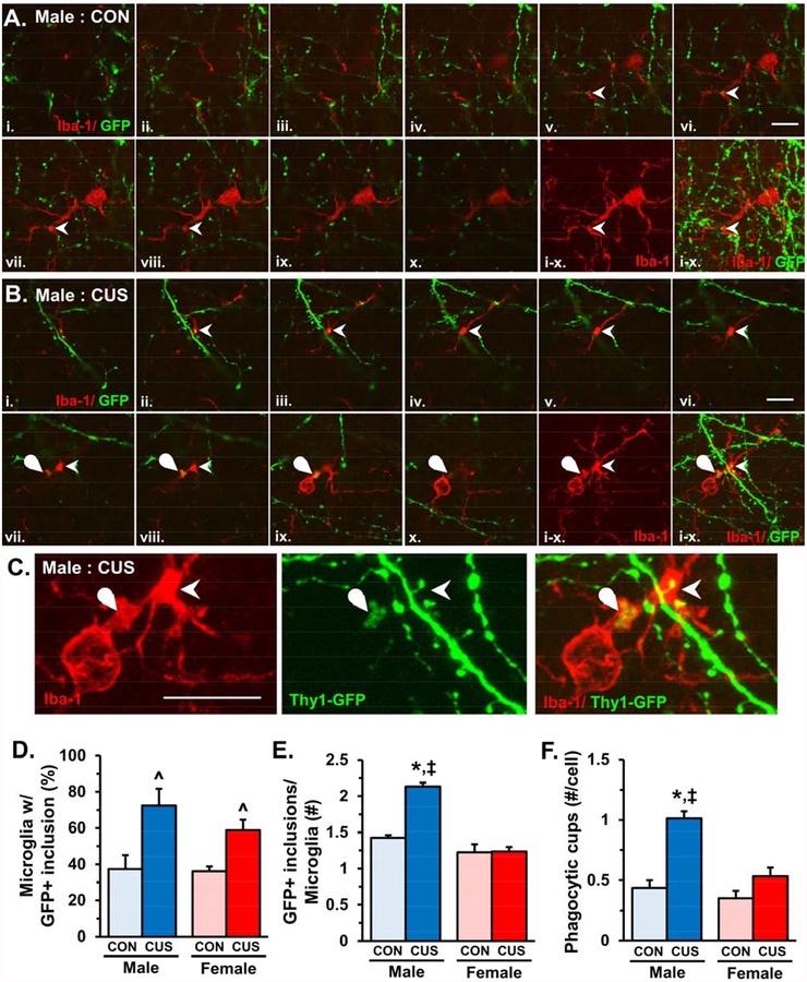
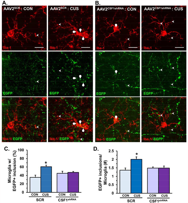
CUS promoted anxiety- and depressive-like behaviors that were associated with increased messenger RNA levels of CSF1 in the PFC. Increased CSF1 messenger RNA levels were also detected in the postmortem dorsolateral PFC of individuals with depression. Moreover, microglia isolated from the frontal cortex of mice exposed to CUS show elevated CSF1 receptor expression and increased phagocytosis of neuronal elements. Notably, functional alterations in microglia were more pronounced in male mice compared with female mice. These functional changes in microglia corresponded with reduced dendritic spine density on pyramidal neurons in layer 1 of the medial PFC. Viral-mediated knockdown of neuronal CSF1 in the medial PFC attenuated microglia-mediated neuronal remodeling and prevented behavioral deficits caused by CUS.
These findings revealed that stress-induced elevations in neuronal CSF1 provokes microglia-mediated neuronal remodeling in the medial PFC, contributing to synaptic deficits and development of anxiety- and depressive-like behavior.
慢性应激暴露会导致前额皮质(PFC)中的神经元萎缩和突触缺失,从而导致焦虑和抑郁样行为的发展。同时,PFC 中的小胶质细胞在应激暴露后会发生形态和功能变化,这表明小胶质细胞可能参与了突触缺失引起的行为后果。
雄性和雌性小鼠接受慢性不可预测应激(CUS)处理,以研究 PFC 中神经元-小胶质细胞相互作用在焦虑和抑郁样行为发展中的作用。使用 Thy1-GFP-M 小鼠评估 PFC 中微胶质细胞介导的神经元重塑和树突棘密度。病毒介导的神经元集落刺激因子 1(CSF1)敲低用于调节 CUS 后小胶质细胞功能和行为后果。
CUS 促进了焦虑和抑郁样行为,这些行为与 PFC 中 CSF1 的信使 RNA 水平升高有关。在患有抑郁症的个体的背外侧 PFC 中也检测到 CSF1 信使 RNA 水平增加。此外,从小鼠 PFC 中分离的小胶质细胞暴露于 CUS 后表现出 CSF1 受体表达增加和神经元成分的吞噬作用增强。值得注意的是,与雌性小鼠相比,雄性小鼠中小胶质细胞的功能改变更为明显。小胶质细胞的这些功能变化与 PFC 内 1 层锥体神经元树突棘密度降低有关。PFC 中的神经元 CSF1 的病毒介导敲低可减弱小胶质细胞介导的神经元重塑,并防止 CUS 引起的行为缺陷。
这些发现表明,应激诱导的神经元 CSF1 升高引发了 PFC 中小胶质细胞介导的神经元重塑,导致突触缺失和焦虑和抑郁样行为的发展。