• 文献检索
  • 文档翻译
  • 深度研究
  • 学术资讯
  • Suppr Zotero 插件Zotero 插件
  • 邀请有礼
  • 套餐&价格
  • 历史记录
应用&插件
Suppr Zotero 插件Zotero 插件浏览器插件Mac 客户端Windows 客户端微信小程序
定价
高级版会员购买积分包购买API积分包
服务
文献检索文档翻译深度研究API 文档MCP 服务
关于我们
关于 Suppr公司介绍联系我们用户协议隐私条款
关注我们

Suppr 超能文献

核心技术专利:CN118964589B侵权必究
粤ICP备2023148730 号-1Suppr @ 2026

文献检索

告别复杂PubMed语法,用中文像聊天一样搜索,搜遍4000万医学文献。AI智能推荐,让科研检索更轻松。

立即免费搜索

文件翻译

保留排版,准确专业,支持PDF/Word/PPT等文件格式,支持 12+语言互译。

免费翻译文档

深度研究

AI帮你快速写综述,25分钟生成高质量综述,智能提取关键信息,辅助科研写作。

立即免费体验

我们真的需要将间充质干细胞分化为胰岛素生成细胞来减轻1型糖尿病中的自身免疫反应吗:胰岛素生成细胞前体的免疫预防作用。

Do we really need to differentiate mesenchymal stem cells into insulin-producing cells for attenuation of the autoimmune responses in type 1 diabetes: immunoprophylactic effects of precursors to insulin-producing cells.

作者信息

Sharma Anshu, Rani Rajni

机构信息

Molecular Immunogenetics Group, National Institute of Immunology, New Delhi, 110067, India.

Systems Biology Group, CSIR-Institute of Genomics and Integrative Biology, New Delhi, 110025, India.

出版信息

Stem Cell Res Ther. 2017 Jul 12;8(1):167. doi: 10.1186/s13287-017-0615-1.

DOI:10.1186/s13287-017-0615-1
PMID:28701182
原文链接:https://pmc.ncbi.nlm.nih.gov/articles/PMC5508489/
Abstract

BACKGROUND

Type 1 diabetes (T1D) is a multifactorial autoimmune disorder where pancreatic beta cells are lost before the clinical manifestations of the disease. Administration of mesenchymal stem cells (MSCs) or MSCs differentiated into insulin-producing cells (IPCs) have yielded limited success when used therapeutically. We have evaluated the immunoprophylactic potentials of precursors to insulin-producing cells (pIPCs) and IPCs in nonobese diabetic (NOD) mice to ask a basic question: do we need to differentiate MSCs into IPCs or will pIPCs suffice to attenuate autoimmune responses in T1D?

METHODS

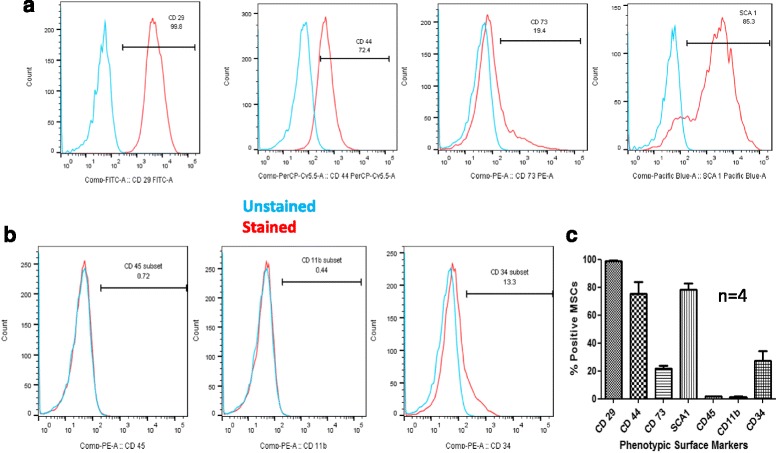
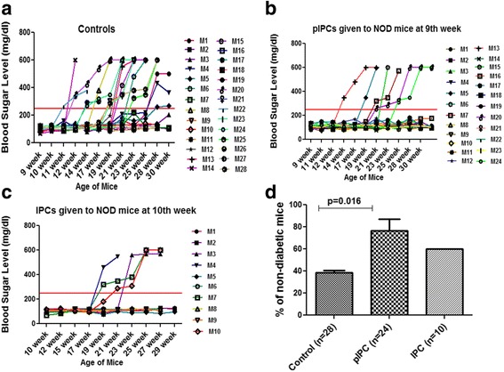
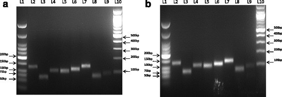
Bone marrow-derived MSCs from Balb/c mice were characterized following the International Society for Cellular Therapy (ISCT) guidelines. MSCs cultured in high-glucose media for 11 to 13 passages were characterized for the expression of pancreatic lineage genes using real-time polymerase chain reaction. Expression of the PDX1 gene in pIPCs was assessed using Western blot and fluorescence-activated cell sorting (FACS). Triple-positive MSCs were differentiated into IPCs using a three-step protocol after sorting them for cell surface markers, i.e. CD29, CD44, and SCA-1. Nonobese diabetic mice were administered pIPCs, IPCs, or phosphate-buffered saline (PBS) into the tail vein at weeks 9 or 10 and followed-up for 29-30 weeks for fasting blood glucose levels. Two consecutive blood sugar levels of more than 250 mg/dl were considered diabetic.

RESULTS

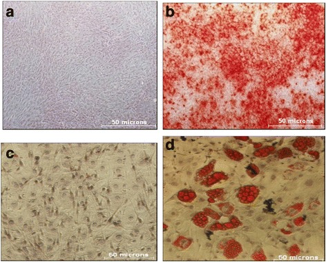
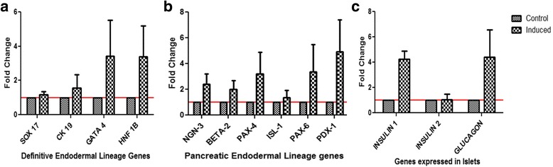
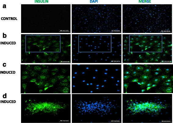
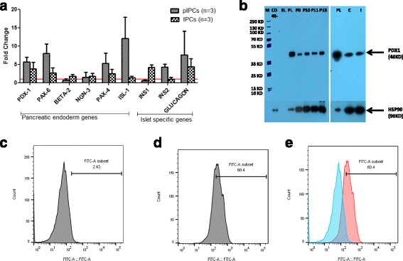
MSCs grown in high-glucose media for 11 to 13 passages expressed genes of the pancreatic lineage such as PDX1, beta2, neurogenin, PAX4, Insulin, and glucagon. Furthermore, Western blot and FACS analysis for PDX-1, a transcription factor necessary for beta cell maturation, confirmed that these cells were precursors of insulin-producing cells (pIPCs). NOD mice administered with pIPCs were better protected from developing diabetes with a protective efficacy of 78.4% (p < 0.009); however, administration of IPCs gave protective efficacy of 55% at the end of 28-30 weeks.

CONCLUSIONS

Precursors to insulin-producing cells seem to have better potential to arrest autoimmune response in type 1 diabetes when administered before the onset of the disease in NOD mice. When translated to humans, autologous mesenchymal stem cells grown in high-glucose media for 10 to 13 passages may have beneficial effects in individuals at high risk of developing type 1 diabetes.

摘要

背景

1型糖尿病(T1D)是一种多因素自身免疫性疾病,在疾病临床表现出现之前胰腺β细胞就已丧失。间充质干细胞(MSCs)或分化为胰岛素产生细胞(IPCs)的MSCs在治疗应用时取得的成功有限。我们评估了胰岛素产生细胞前体(pIPCs)和IPCs在非肥胖糖尿病(NOD)小鼠中的免疫预防潜力,以探讨一个基本问题:我们是否需要将MSCs分化为IPCs,还是pIPCs就足以减弱T1D中的自身免疫反应?

方法

按照国际细胞治疗协会(ISCT)指南对来自Balb/c小鼠的骨髓源性MSCs进行鉴定。使用实时聚合酶链反应对在高糖培养基中培养11至13代的MSCs进行胰腺谱系基因表达的鉴定。使用蛋白质免疫印迹法和荧光激活细胞分选(FACS)评估pIPCs中PDX1基因的表达。对细胞表面标志物即CD29、CD44和SCA-1进行分选后,采用三步方案将三阳性MSCs分化为IPCs。在第9周或第10周给非肥胖糖尿病小鼠尾静脉注射pIPCs、IPCs或磷酸盐缓冲盐水(PBS),并随访29 - 30周以检测空腹血糖水平。连续两次血糖水平超过250mg/dl被视为糖尿病。

结果

在高糖培养基中培养11至13代的MSCs表达胰腺谱系基因,如PDX1、β2、神经生成素、PAX4、胰岛素和胰高血糖素。此外,对β细胞成熟所必需的转录因子PDX - 1进行的蛋白质免疫印迹法和FACS分析证实这些细胞是胰岛素产生细胞前体(pIPCs)。接受pIPCs治疗的NOD小鼠对糖尿病的发展具有更好的保护作用,保护效力为78.4%(p < 0.009);然而,在28 - 30周结束时,给予IPCs的保护效力为55%。

结论

在NOD小鼠疾病发作前给予胰岛素产生细胞前体似乎在阻止1型糖尿病自身免疫反应方面具有更好的潜力。如果应用于人类,在高糖培养基中培养10至13代的自体间充质干细胞可能对有发展为1型糖尿病高风险的个体具有有益作用。

https://cdn.ncbi.nlm.nih.gov/pmc/blobs/95f1/5508489/8b6ba2aabac1/13287_2017_615_Fig7_HTML.jpg
https://cdn.ncbi.nlm.nih.gov/pmc/blobs/95f1/5508489/bb803a4b5743/13287_2017_615_Fig1_HTML.jpg
https://cdn.ncbi.nlm.nih.gov/pmc/blobs/95f1/5508489/133647094d40/13287_2017_615_Fig2_HTML.jpg
https://cdn.ncbi.nlm.nih.gov/pmc/blobs/95f1/5508489/624e3370ea2e/13287_2017_615_Fig3_HTML.jpg
https://cdn.ncbi.nlm.nih.gov/pmc/blobs/95f1/5508489/02d006d41aa8/13287_2017_615_Fig4_HTML.jpg
https://cdn.ncbi.nlm.nih.gov/pmc/blobs/95f1/5508489/869386c8e8cf/13287_2017_615_Fig5_HTML.jpg
https://cdn.ncbi.nlm.nih.gov/pmc/blobs/95f1/5508489/1ebdb9a3b246/13287_2017_615_Fig6_HTML.jpg
https://cdn.ncbi.nlm.nih.gov/pmc/blobs/95f1/5508489/8b6ba2aabac1/13287_2017_615_Fig7_HTML.jpg

相似文献

1
Do we really need to differentiate mesenchymal stem cells into insulin-producing cells for attenuation of the autoimmune responses in type 1 diabetes: immunoprophylactic effects of precursors to insulin-producing cells.我们真的需要将间充质干细胞分化为胰岛素生成细胞来减轻1型糖尿病中的自身免疫反应吗:胰岛素生成细胞前体的免疫预防作用。
Stem Cell Res Ther. 2017 Jul 12;8(1):167. doi: 10.1186/s13287-017-0615-1.
2
Undifferentiated Wharton's Jelly Mesenchymal Stem Cell Transplantation Induces Insulin-Producing Cell Differentiation and Suppression of T-Cell-Mediated Autoimmunity in Nonobese Diabetic Mice.未分化的沃顿胶间充质干细胞移植诱导非肥胖糖尿病小鼠产生胰岛素分泌细胞并抑制T细胞介导的自身免疫反应。
Cell Transplant. 2015;24(8):1555-70. doi: 10.3727/096368914X683016. Epub 2014 Jul 15.
3
Mesenchymal stem cells and differentiated insulin producing cells are new horizons for pancreatic regeneration in type I diabetes mellitus.间充质干细胞和分化的胰岛素产生细胞是I型糖尿病胰腺再生的新方向。
Int J Biochem Cell Biol. 2017 Jun;87:77-85. doi: 10.1016/j.biocel.2017.03.018. Epub 2017 Apr 3.
4
Transplantation of differentiated umbilical cord mesenchymal cells under kidney capsule for control of type I diabetes in rat.将分化的脐带间充质细胞移植到大鼠肾被膜下以控制I型糖尿病
Tissue Cell. 2015 Aug;47(4):395-405. doi: 10.1016/j.tice.2015.04.008. Epub 2015 May 21.
5
Insulin-Producing Cells Differentiated from Human Bone Marrow Mesenchymal Stem Cells In Vitro Ameliorate Streptozotocin-Induced Diabetic Hyperglycemia.体外从人骨髓间充质干细胞分化而来的胰岛素生成细胞可改善链脲佐菌素诱导的糖尿病性高血糖症。
PLoS One. 2016 Jan 12;11(1):e0145838. doi: 10.1371/journal.pone.0145838. eCollection 2016.
6
Transplantation of insulin-producing cells differentiated from human periosteum-derived progenitor cells ameliorate hyperglycemia in diabetic mice.人骨膜来源祖细胞诱导分化的胰岛素分泌细胞移植改善糖尿病小鼠的高血糖。
Transplantation. 2014 Nov 27;98(10):1040-7. doi: 10.1097/TP.0000000000000388.
7
Multipotent mesenchymal stromal cells from patients with newly diagnosed type 1 diabetes mellitus exhibit preserved in vitro and in vivo immunomodulatory properties.新诊断的1型糖尿病患者的多能间充质基质细胞在体外和体内均表现出保留的免疫调节特性。
Stem Cell Res Ther. 2016 Jan 18;7:14. doi: 10.1186/s13287-015-0261-4.
8
Differentiation of bone marrow-derived mesenchymal stem cells from diabetic patients into insulin-producing cells in vitro.体外将糖尿病患者骨髓来源的间充质干细胞分化为胰岛素分泌细胞。
Chin Med J (Engl). 2007 May 5;120(9):771-6.
9
Differentiation of Human Bone Marrow-Derived Mesenchymal Stem Cells into Insulin-Producing Cells: Evidence for Further Maturation In Vivo.人骨髓间充质干细胞向胰岛素分泌细胞的分化:体内进一步成熟的证据
Biomed Res Int. 2015;2015:575837. doi: 10.1155/2015/575837. Epub 2015 May 12.
10
HCELL Expression on Murine MSC Licenses Pancreatotropism and Confers Durable Reversal of Autoimmune Diabetes in NOD Mice.小鼠间充质干细胞上的HCELL表达赋予胰腺趋向性并持久逆转NOD小鼠的自身免疫性糖尿病。
Stem Cells. 2015 May;33(5):1523-31. doi: 10.1002/stem.1948.

引用本文的文献

1
Differentiation of Alginate-Encapsulated Wharton Jelly-Derived Mesenchymal Stem Cells into Insulin Producing Cells.藻酸盐包封的脐带华通氏胶间充质干细胞向胰岛素产生细胞的分化
Cell J. 2022 Aug 28;24(8):449-457. doi: 10.22074/cellj.2022.8081.
2
Mesenchymal Stem Cells: An Excellent Candidate for the Treatment of Diabetes Mellitus.间充质干细胞:治疗糖尿病的理想候选者。
Int J Endocrinol. 2021 May 28;2021:9938658. doi: 10.1155/2021/9938658. eCollection 2021.
3
Advances and complications of regenerative medicine in diabetes therapy.

本文引用的文献

1
Mesenchymal Stem Cells: Rising Concerns over Their Application in Treatment of Type One Diabetes Mellitus.间充质干细胞:对其在1型糖尿病治疗中应用的日益关注。
J Diabetes Res. 2015;2015:675103. doi: 10.1155/2015/675103. Epub 2015 Oct 20.
2
Immunomodulatory characteristics of mesenchymal stem cells and their role in the treatment of multiple sclerosis.间充质干细胞的免疫调节特性及其在多发性硬化症治疗中的作用。
Cell Immunol. 2015 Feb;293(2):113-21. doi: 10.1016/j.cellimm.2015.01.002. Epub 2015 Jan 10.
3
Three-dimensional differentiation of bone marrow-derived mesenchymal stem cells into insulin-producing cells.
再生医学在糖尿病治疗中的进展与并发症
PeerJ. 2020 Sep 8;8:e9746. doi: 10.7717/peerj.9746. eCollection 2020.
4
Direct lineage tracing reveals Activin-a potential for improved pancreatic homing of bone marrow mesenchymal stem cells and efficient ß-cell regeneration in vivo.直接谱系追踪显示激活素 A 可提高骨髓间充质干细胞对胰腺的归巢能力,并在体内有效促进β细胞再生。
Stem Cell Res Ther. 2020 Jul 30;11(1):327. doi: 10.1186/s13287-020-01843-z.
5
Targeted induction of bone marrow mesenchymal stem cells to have effectiveness on diabetic pancreatic restoration.靶向诱导骨髓间充质干细胞对糖尿病胰腺修复的有效性。
In Vitro Cell Dev Biol Anim. 2019 Jun;55(6):453-461. doi: 10.1007/s11626-019-00364-1. Epub 2019 May 28.
6
Stem cells as a potential therapy for diabetes mellitus: a call-to-action in Latin America.干细胞作为糖尿病的一种潜在疗法:拉丁美洲的行动呼吁。
Diabetol Metab Syndr. 2019 Feb 18;11:20. doi: 10.1186/s13098-019-0415-0. eCollection 2019.
7
Human urine-derived stem cells play a novel role in the treatment of STZ-induced diabetic mice.人尿源干细胞在 STZ 诱导的糖尿病小鼠治疗中发挥新作用。
J Mol Histol. 2018 Aug;49(4):419-428. doi: 10.1007/s10735-018-9772-5. Epub 2018 Apr 19.
骨髓间充质干细胞向胰岛素分泌细胞的三维分化
Tissue Cell. 2015 Feb;47(1):66-72. doi: 10.1016/j.tice.2014.11.005. Epub 2014 Dec 5.
4
Islet cell transplantation for the treatment of type 1 diabetes: recent advances and future challenges.胰岛细胞移植治疗 1 型糖尿病:最新进展与未来挑战。
Diabetes Metab Syndr Obes. 2014 Jun 23;7:211-23. doi: 10.2147/DMSO.S50789. eCollection 2014.
5
The life and fate of mesenchymal stem cells.间充质干细胞的生活与命运。
Front Immunol. 2014 May 19;5:148. doi: 10.3389/fimmu.2014.00148. eCollection 2014.
6
In vitro hepatic trans-differentiation of human mesenchymal stem cells using sera from congestive/ischemic liver during cardiac failure.在心力衰竭期间使用充血/缺血性肝血清体外诱导人骨髓间充质干细胞向肝系细胞转分化。
PLoS One. 2014 Mar 18;9(3):e92397. doi: 10.1371/journal.pone.0092397. eCollection 2014.
7
Human adipose-derived mesenchymal stem cells: a better cell source for nervous system regeneration.人脂肪间充质干细胞:神经系统再生的更佳细胞来源。
Chin Med J (Engl). 2014;127(2):329-37.
8
Intraportal injection of insulin-producing cells generated from human bone marrow mesenchymal stem cells decreases blood glucose level in diabetic rats.经人骨髓间充质干细胞诱导分化的胰岛素分泌细胞门静脉内注射可降低糖尿病大鼠的血糖水平。
Endocr Res. 2014;39(1):26-33. doi: 10.3109/07435800.2013.797432. Epub 2013 Jun 17.
9
Effect of combined therapy of human Wharton's jelly-derived mesenchymal stem cells from umbilical cord with sitagliptin in type 2 diabetic rats.脐带人羊膜间充质干细胞与西他列汀联合治疗对2型糖尿病大鼠的影响。
Endocrine. 2014 Mar;45(2):279-87. doi: 10.1007/s12020-013-9984-0. Epub 2013 May 18.
10
Reprogramming of bone marrow-derived mesenchymal stem cells into functional insulin-producing cells by chemical regimen.通过化学方案将骨髓间充质干细胞重编程为功能性胰岛素分泌细胞。
Am J Stem Cells. 2012 Mar 28;1(2):128-37. Print 2012.