• 文献检索
  • 文档翻译
  • 深度研究
  • 学术资讯
  • Suppr Zotero 插件Zotero 插件
  • 邀请有礼
  • 套餐&价格
  • 历史记录
应用&插件
Suppr Zotero 插件Zotero 插件浏览器插件Mac 客户端Windows 客户端微信小程序
定价
高级版会员购买积分包购买API积分包
服务
文献检索文档翻译深度研究API 文档MCP 服务
关于我们
关于 Suppr公司介绍联系我们用户协议隐私条款
关注我们

Suppr 超能文献

核心技术专利:CN118964589B侵权必究
粤ICP备2023148730 号-1Suppr @ 2026

文献检索

告别复杂PubMed语法,用中文像聊天一样搜索,搜遍4000万医学文献。AI智能推荐,让科研检索更轻松。

立即免费搜索

文件翻译

保留排版,准确专业,支持PDF/Word/PPT等文件格式,支持 12+语言互译。

免费翻译文档

深度研究

AI帮你快速写综述,25分钟生成高质量综述,智能提取关键信息,辅助科研写作。

立即免费体验

II级和III级胶质瘤的扩散峰度成像——一项关于临床症状下瘤周肿瘤浸润、肿瘤分级及亚型的研究

Diffusion Kurtosis Imaging of Gliomas Grades II and III - A Study of Perilesional Tumor Infiltration, Tumor Grades and Subtypes at Clinical Presentation.

作者信息

Delgado Anna F, Fahlström Markus, Nilsson Markus, Berntsson Shala G, Zetterling Maria, Libard Sylwia, Alafuzoff Irina, van Westen Danielle, Lätt Jimmy, Smits Anja, Larsson Elna-Marie

机构信息

Department of Surgical Sciences, Radiology, Uppsala University, Uppsala, Sweden.

Department of Clinical Neuroscience, Karolinska Institute, Stockholm, Sweden.

出版信息

Radiol Oncol. 2017 Feb 15;51(2):121-129. doi: 10.1515/raon-2017-0010. eCollection 2017 Jun.

DOI:10.1515/raon-2017-0010
PMID:28740446
原文链接:https://pmc.ncbi.nlm.nih.gov/articles/PMC5514651/
Abstract

BACKGROUND

Diffusion kurtosis imaging (DKI) allows for assessment of diffusion influenced by microcellular structures. We analyzed DKI in suspected low-grade gliomas prior to histopathological diagnosis. The aim was to investigate if diffusion parameters in the perilesional normal-appearing white matter (NAWM) differed from contralesional white matter, and to investigate differences between glioma malignancy grades II and III and glioma subtypes (astrocytomas and oligodendrogliomas).

PATIENTS AND METHODS

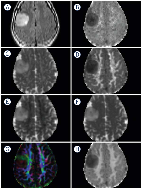
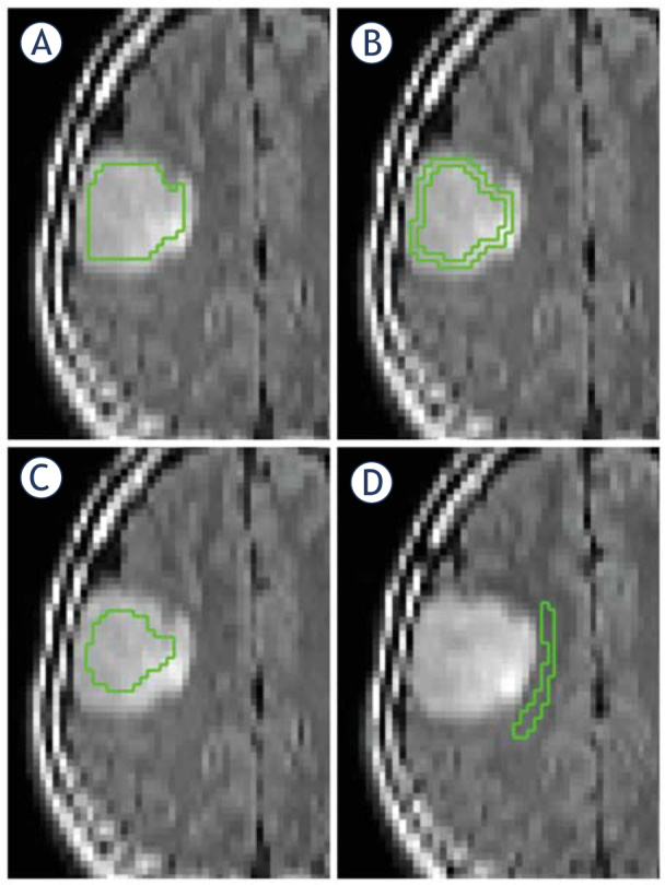
Forty-eight patients with suspected low-grade glioma were prospectively recruited to this institutional review board-approved study and investigated with preoperative DKI at 3T after written informed consent. Patients with histologically proven glioma grades II or III were further analyzed (n=35). Regions of interest (ROIs) were delineated on T2FLAIR images and co-registered to diffusion MRI parameter maps. Mean DKI data were compared between perilesional and contralesional NAWM (student's -test for dependent samples, Wilcoxon matched pairs test). Histogram DKI data were compared between glioma types and glioma grades (multiple comparisons of mean ranks for all groups). The discriminating potential for DKI in assessing glioma type and grade was assessed with receiver operating characteristics (ROC) curves.

RESULTS

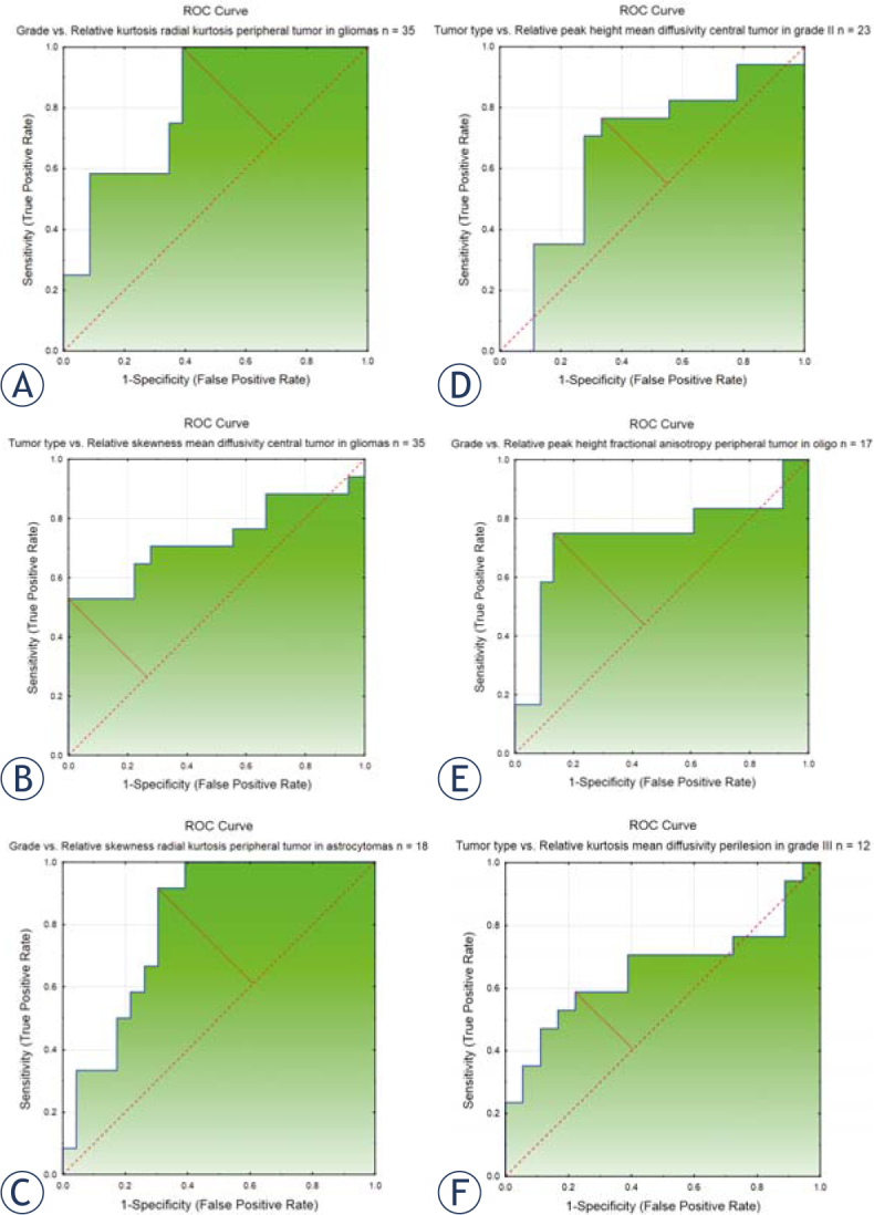
There were significant differences in all mean DKI variables between perilesional and contralesional NAWM (=<0.000), except for axial kurtosis (=0.099). Forty-four histogram variables differed significantly between glioma grades II (n=23) and III (n=12) (p=0.003-0.048) and 10 variables differed significantly between ACs (n=18) and ODs (n=17) (=0.011-0.050). ROC curves of the best discriminating variables had an area under the curve (AUC) of 0.657-0.815.

CONCLUSIONS

Mean DKI variables in perilesional NAWM differ significantly from contralesional NAWM, suggesting altered microstructure by tumor infiltration not depicted on morphological MRI. Histogram analysis of DKI data identifies differences between glioma grades and subtypes.

摘要

背景

扩散峰度成像(DKI)可用于评估受微细胞结构影响的扩散情况。我们在组织病理学诊断之前对疑似低级别胶质瘤进行了DKI分析。目的是研究瘤周正常外观白质(NAWM)中的扩散参数是否与对侧白质不同,以及研究胶质瘤II级和III级以及胶质瘤亚型(星形细胞瘤和少突胶质细胞瘤)之间的差异。

患者与方法

48例疑似低级别胶质瘤患者被前瞻性纳入本机构审查委员会批准的研究,并在签署书面知情同意书后于3T进行术前DKI检查。对组织学证实为II级或III级胶质瘤的患者进行进一步分析(n = 35)。在T2FLAIR图像上勾勒出感兴趣区域(ROI),并将其与扩散MRI参数图进行配准。比较瘤周和对侧NAWM之间的平均DKI数据(配对样本的学生t检验,Wilcoxon配对检验)。比较胶质瘤类型和胶质瘤级别之间的直方图DKI数据(所有组平均秩次的多重比较)。使用受试者工作特征(ROC)曲线评估DKI在评估胶质瘤类型和级别方面的鉴别潜力。

结果

除轴向峰度(p = 0.099)外,瘤周和对侧NAWM之间所有平均DKI变量均存在显著差异(p <= 0.000)。44个直方图变量在胶质瘤II级(n = 23)和III级(n = 12)之间存在显著差异(p = 0.003 - 0.048),10个变量在星形细胞瘤(n = 18)和少突胶质细胞瘤(n = 17)之间存在显著差异(p = 0.011 - 0.050)。最佳鉴别变量的ROC曲线下面积(AUC)为0.657 - 0.815。

结论

瘤周NAWM中的平均DKI变量与对侧NAWM有显著差异,提示肿瘤浸润导致微观结构改变,而形态学MRI未显示。DKI数据的直方图分析可识别胶质瘤级别和亚型之间的差异。

https://cdn.ncbi.nlm.nih.gov/pmc/blobs/2114/5514651/d29c930b8b99/raon-51-121-g003.jpg
https://cdn.ncbi.nlm.nih.gov/pmc/blobs/2114/5514651/5984a2d66af1/raon-51-121-g001.jpg
https://cdn.ncbi.nlm.nih.gov/pmc/blobs/2114/5514651/1f7d8768ca29/raon-51-121-g002.jpg
https://cdn.ncbi.nlm.nih.gov/pmc/blobs/2114/5514651/d29c930b8b99/raon-51-121-g003.jpg
https://cdn.ncbi.nlm.nih.gov/pmc/blobs/2114/5514651/5984a2d66af1/raon-51-121-g001.jpg
https://cdn.ncbi.nlm.nih.gov/pmc/blobs/2114/5514651/1f7d8768ca29/raon-51-121-g002.jpg
https://cdn.ncbi.nlm.nih.gov/pmc/blobs/2114/5514651/d29c930b8b99/raon-51-121-g003.jpg

相似文献

1
Diffusion Kurtosis Imaging of Gliomas Grades II and III - A Study of Perilesional Tumor Infiltration, Tumor Grades and Subtypes at Clinical Presentation.II级和III级胶质瘤的扩散峰度成像——一项关于临床症状下瘤周肿瘤浸润、肿瘤分级及亚型的研究
Radiol Oncol. 2017 Feb 15;51(2):121-129. doi: 10.1515/raon-2017-0010. eCollection 2017 Jun.
2
Histogram analysis of diffusion kurtosis imaging derived maps may distinguish between low and high grade gliomas before surgery.基于扩散峰度成像衍生图的直方图分析可在术前区分低级别和高级别胶质瘤。
Eur Radiol. 2018 Apr;28(4):1748-1755. doi: 10.1007/s00330-017-5108-1. Epub 2017 Nov 16.
3
Histogram analysis of diffusion kurtosis imaging estimates for in vivo assessment of 2016 WHO glioma grades: A cross-sectional observational study.基于体素内不相干运动扩散峰度成像的直方图分析对 2016 年世界卫生组织脑胶质瘤分级的评估:一项横断面观察性研究。
Eur J Radiol. 2017 Oct;95:202-211. doi: 10.1016/j.ejrad.2017.08.008. Epub 2017 Aug 18.
4
Value of diffusion kurtosis imaging in assessing low-grade gliomas.弥散峰度成像在评估低级别胶质瘤中的价值。
J Magn Reson Imaging. 2018 Dec;48(6):1551-1558. doi: 10.1002/jmri.26012. Epub 2018 Mar 23.
5
Gliomas: diffusion kurtosis MR imaging in grading.脑胶质瘤:扩散峰度磁共振成像分级。
Radiology. 2012 May;263(2):492-501. doi: 10.1148/radiol.12110927. Epub 2012 Mar 8.
6
Assessment of tissue heterogeneity using diffusion tensor and diffusion kurtosis imaging for grading gliomas.使用扩散张量成像和扩散峰度成像评估组织异质性以对胶质瘤进行分级。
Neuroradiology. 2016 Dec;58(12):1217-1231. doi: 10.1007/s00234-016-1758-y. Epub 2016 Oct 29.
7
Glioma Type Prediction with Dynamic Contrast-Enhanced MR Imaging and Diffusion Kurtosis Imaging-A Standardized Multicenter Study.基于动态对比增强磁共振成像和扩散峰度成像的胶质瘤类型预测——一项标准化多中心研究
Cancers (Basel). 2024 Jul 25;16(15):2644. doi: 10.3390/cancers16152644.
8
Tumor Multiregional Mean Apparent Propagator (MAP) Features in Evaluating Gliomas-A Comparative Study With Diffusion Kurtosis Imaging (DKI).肿瘤多区域平均表观扩散系数(MAP)特征在胶质瘤评估中的应用——与扩散峰度成像(DKI)的比较研究。
J Magn Reson Imaging. 2024 Oct;60(4):1532-1546. doi: 10.1002/jmri.29202. Epub 2023 Dec 22.
9
Cerebral gliomas: diffusional kurtosis imaging analysis of microstructural differences.脑胶质瘤:扩散峰度成像分析的微观结构差异。
Radiology. 2010 Mar;254(3):876-81. doi: 10.1148/radiol.09090819. Epub 2010 Jan 20.
10
Diagnostic performance between MR amide proton transfer (APT) and diffusion kurtosis imaging (DKI) in glioma grading and IDH mutation status prediction at 3 T.3T 下磁共振酰胺质子转移(APT)和扩散峰度成像(DKI)在胶质瘤分级和 IDH 突变状态预测中的诊断性能。
Eur J Radiol. 2021 Jan;134:109466. doi: 10.1016/j.ejrad.2020.109466. Epub 2020 Dec 3.

引用本文的文献

1
Individual Brain Tumor Invasion Mapping Based on Diffusion Kurtosis Imaging.基于扩散峰度成像的个体脑肿瘤侵袭图谱
Sovrem Tekhnologii Med. 2025;17(1):81-90. doi: 10.17691/stm2025.17.1.08. Epub 2025 Feb 28.
2
Enhancing Diagnostic Precision: Evaluation of Preprocessing Filters in Simple Diffusion Kurtosis Imaging for Head and Neck Tumors.提高诊断精度:头颈部肿瘤简单扩散峰度成像中预处理滤波器的评估
J Clin Med. 2024 Mar 20;13(6):1783. doi: 10.3390/jcm13061783.
3
A comparative study of diffusion kurtosis imaging and diffusion tensor imaging in detecting corticospinal tract impairment in diffuse glioma patients.

本文引用的文献

1
The link between diffusion MRI and tumor heterogeneity: Mapping cell eccentricity and density by diffusional variance decomposition (DIVIDE).扩散加权磁共振成像与肿瘤异质性之间的联系:通过扩散方差分解(DIVIDE)绘制细胞偏心率和密度图。
Neuroimage. 2016 Nov 15;142:522-532. doi: 10.1016/j.neuroimage.2016.07.038. Epub 2016 Jul 20.
2
The 2016 World Health Organization Classification of Tumors of the Central Nervous System: a summary.2016 年世界卫生组织中枢神经系统肿瘤分类:概述。
Acta Neuropathol. 2016 Jun;131(6):803-20. doi: 10.1007/s00401-016-1545-1. Epub 2016 May 9.
3
Extension of diffuse low-grade gliomas beyond radiological borders as shown by the coregistration of histopathological and magnetic resonance imaging data.
弥散峰度成像与弥散张量成像在检测弥漫性胶质瘤患者皮质脊髓束损伤中的对比研究。
Neuroradiology. 2024 May;66(5):785-796. doi: 10.1007/s00234-024-03332-z. Epub 2024 Mar 13.
4
Perifocal Zone of Brain Gliomas: Application of Diffusion Kurtosis and Perfusion MRI Values for Tumor Invasion Border Determination.脑胶质瘤的瘤周区域:扩散峰度成像与灌注磁共振成像值在肿瘤浸润边界确定中的应用
Cancers (Basel). 2023 May 15;15(10):2760. doi: 10.3390/cancers15102760.
5
Brain Diffusion Weighted Imaging Study of Mongolian Idiopathic Epilepsy.蒙古特发性癫痫的脑弥散加权成像研究。
J Healthc Eng. 2022 Nov 28;2022:6978116. doi: 10.1155/2022/6978116. eCollection 2022.
6
Challenges and opportunities for advanced neuroimaging of glioblastoma.胶质母细胞瘤的高级神经影像学的挑战与机遇。
Br J Radiol. 2023 Jan 1;96(1141):20211232. doi: 10.1259/bjr.20211232. Epub 2022 Oct 14.
7
Mapping early tumor response to radiotherapy using diffusion kurtosis imaging*.使用扩散峰度成像*对放疗早期肿瘤反应进行成像。
Neuroradiol J. 2023 Apr;36(2):198-205. doi: 10.1177/19714009221122204. Epub 2022 Aug 24.
8
Two Patterns of White Matter Connection in Multiple Gliomas: Evidence from Probabilistic Fiber Tracking.多发性胶质瘤中白质连接的两种模式:来自概率纤维追踪的证据
J Clin Med. 2022 Jun 27;11(13):3693. doi: 10.3390/jcm11133693.
9
Machine Learning Based on Diffusion Kurtosis Imaging Histogram Parameters for Glioma Grading.基于扩散峰度成像直方图参数的机器学习用于脑胶质瘤分级
J Clin Med. 2022 Apr 21;11(9):2310. doi: 10.3390/jcm11092310.
10
Sensitivity of Diffusion MRI to White Matter Pathology: Influence of Diffusion Protocol, Magnetic Field Strength, and Processing Pipeline in Systemic Lupus Erythematosus.扩散加权磁共振成像对脑白质病变的敏感性:扩散协议、磁场强度及处理流程在系统性红斑狼疮中的影响
Front Neurol. 2022 Apr 26;13:837385. doi: 10.3389/fneur.2022.837385. eCollection 2022.
弥散性低级别胶质瘤的影像学边界外侵犯:基于组织病理学和磁共振成像数据的配准。
J Neurosurg. 2016 Nov;125(5):1155-1166. doi: 10.3171/2015.10.JNS15583. Epub 2016 Feb 26.
4
Diffusion kurtosis imaging can efficiently assess the glioma grade and cellular proliferation.扩散峰度成像能够有效地评估胶质瘤的分级和细胞增殖情况。
Oncotarget. 2015 Dec 8;6(39):42380-93. doi: 10.18632/oncotarget.5675.
5
Neurological Impairment Linked with Cortico-Subcortical Infiltration of Diffuse Low-Grade Gliomas at Initial Diagnosis Supports Early Brain Plasticity.初诊时弥漫性低级别胶质瘤的皮质-皮质下浸润与神经功能损害相关,这支持早期脑可塑性。
Front Neurol. 2015 Jun 10;6:137. doi: 10.3389/fneur.2015.00137. eCollection 2015.
6
Mean Diffusional Kurtosis in Patients with Glioma: Initial Results with a Fast Imaging Method in a Clinical Setting.胶质瘤患者的平均扩散峰度:临床环境中快速成像方法的初步结果
AJNR Am J Neuroradiol. 2015 Aug;36(8):1472-8. doi: 10.3174/ajnr.A4311. Epub 2015 May 14.
7
Dynamic Contrast-Enhanced Perfusion MRI and Diffusion-Weighted Imaging in Grading of Gliomas.动态对比增强灌注磁共振成像与扩散加权成像在胶质瘤分级中的应用
J Neuroimaging. 2015 Sep-Oct;25(5):792-8. doi: 10.1111/jon.12239. Epub 2015 Apr 13.
8
Direct Cranial Nerve Involvement by Gliomas: Case Series and Review of the Literature.神经胶质瘤直接侵犯脑神经:病例系列及文献综述
AJNR Am J Neuroradiol. 2015 Jul;36(7):1349-54. doi: 10.3174/ajnr.A4287. Epub 2015 Apr 9.
9
Discrimination between glioma grades II and III in suspected low-grade gliomas using dynamic contrast-enhanced and dynamic susceptibility contrast perfusion MR imaging: a histogram analysis approach.利用动态对比增强和动态磁敏感对比灌注磁共振成像,通过直方图分析方法鉴别疑似低级别胶质瘤中的二级和三级胶质瘤。
Neuroradiology. 2014 Dec;56(12):1031-8. doi: 10.1007/s00234-014-1426-z. Epub 2014 Sep 10.
10
Machine learning in preoperative glioma MRI: survival associations by perfusion-based support vector machine outperforms traditional MRI.术前胶质瘤磁共振成像中的机器学习:基于灌注的支持向量机的生存关联优于传统磁共振成像。
J Magn Reson Imaging. 2014 Jul;40(1):47-54. doi: 10.1002/jmri.24390. Epub 2013 Nov 13.