• 文献检索
  • 文档翻译
  • 深度研究
  • 学术资讯
  • Suppr Zotero 插件Zotero 插件
  • 邀请有礼
  • 套餐&价格
  • 历史记录
应用&插件
Suppr Zotero 插件Zotero 插件浏览器插件Mac 客户端Windows 客户端微信小程序
定价
高级版会员购买积分包购买API积分包
服务
文献检索文档翻译深度研究API 文档MCP 服务
关于我们
关于 Suppr公司介绍联系我们用户协议隐私条款
关注我们

Suppr 超能文献

核心技术专利:CN118964589B侵权必究
粤ICP备2023148730 号-1Suppr @ 2026

文献检索

告别复杂PubMed语法,用中文像聊天一样搜索,搜遍4000万医学文献。AI智能推荐,让科研检索更轻松。

立即免费搜索

文件翻译

保留排版,准确专业,支持PDF/Word/PPT等文件格式,支持 12+语言互译。

免费翻译文档

深度研究

AI帮你快速写综述,25分钟生成高质量综述,智能提取关键信息,辅助科研写作。

立即免费体验

磁共振成像减少了乳腺癌患者肿瘤床无手术夹时临床靶区轮廓勾画的差异,以提高靶区勾画准确性。

MRI Reduces Variation of Contouring for Boost Clinical Target Volume in Breast Cancer Patients Without Surgical Clips in the Tumour Bed.

作者信息

Al-Hammadi Noora, Caparrotti Palmira, Divakar Saju, Riyas Mohamed, Chandramouli Suparna Halsnad, Hammoud Rabih, Hayes Jillian, Mc Garry Maeve, Paloor Satheesh Prasad, Petric Primoz

机构信息

Department of Radiation Oncology, National Center for Cancer Care and Research, Hamad Medical Corporation, Doha, Qatar.

出版信息

Radiol Oncol. 2017 Mar 17;51(2):160-168. doi: 10.1515/raon-2017-0014. eCollection 2017 Jun.

DOI:10.1515/raon-2017-0014
PMID:28740451
原文链接:https://pmc.ncbi.nlm.nih.gov/articles/PMC5514656/
Abstract

BACKGROUND

Omitting the placement of clips inside tumour bed during breast cancer surgery poses a challenge for delineation of lumpectomy cavity clinical target volume (CTV). We aimed to quantify inter-observer variation and accuracy for CT- and MRI-based segmentation of CTV in patients without clips.

PATIENTS AND METHODS

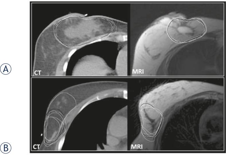
CT- and MRI-simulator images of 12 breast cancer patients, treated by breast conserving surgery and radiotherapy, were included in this study. Five radiation oncologists recorded the cavity visualization score (CVS) and delineated CTV on both modalities. Expert-consensus (EC) contours were delineated by a senior radiation oncologist, respecting opinions of all observers. Inter-observer volumetric variation and generalized conformity index (CI) were calculated. Deviations from EC contour were quantified by the accuracy index (AI) and inter-delineation distances (IDD).

RESULTS

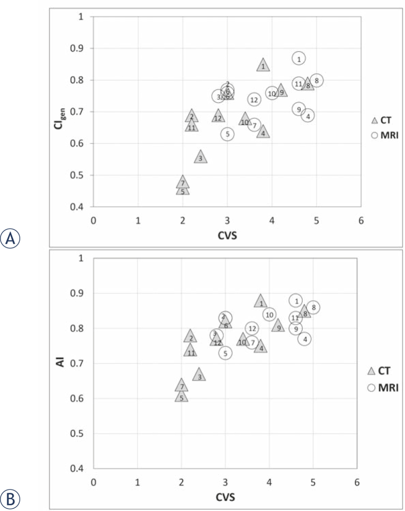
Mean CVS was 3.88 +/- 0.99 and 3.05 +/- 1.07 for MRI and CT, respectively (p = 0.001). Mean volumes of CTV were similar: 154 +/- 26 cm on CT and 152 +/- 19 cm on MRI. Mean CI and AI were superior for MRI when compared with CT (CI: 0.74 +/- 0.07 vs. 0.67 +/- 0.12, p = 0.007; AI: 0.81 +/- 0.04 vs. 0.76 +/- 0.07; p = 0.004). CI and AI increased with increasing CVS. Mean IDD was 3 mm +/- 1.5 mm and 3.6 mm +/- 2.3 mm for MRI and CT, respectively (p = 0.017).

CONCLUSIONS

When compared with CT, MRI improved visualization of post-lumpectomy changes, reduced interobserver variation and improved the accuracy of CTV contouring in patients without clips in the tumour bed. Further studies with bigger sample sizes are needed to confirm our findings.

摘要

背景

在乳腺癌手术中不放置夹子于瘤床内,给确定保乳手术腔临床靶区(CTV)带来了挑战。我们旨在量化在无夹子患者中基于CT和MRI的CTV分割的观察者间差异及准确性。

患者与方法

本研究纳入了12例接受保乳手术和放疗的乳腺癌患者的CT和MRI模拟图像。5名放射肿瘤学家记录了两种模态下的腔可视化评分(CVS)并勾画CTV。由一名资深放射肿瘤学家勾画专家共识(EC)轮廓,参考所有观察者的意见。计算观察者间体积差异和广义符合指数(CI)。通过准确性指数(AI)和轮廓间距离(IDD)量化与EC轮廓的偏差。

结果

MRI和CT的平均CVS分别为3.88±0.99和3.05±1.07(p = 0.001)。CTV的平均体积相似:CT上为154±26 cm,MRI上为152±19 cm。与CT相比,MRI的平均CI和AI更优(CI:0.74±0.07对0.67±0.12,p = 0.007;AI:0.81±0.04对0.76±0.07;p = 0.004)。CI和AI随CVS增加而升高。MRI和CT的平均IDD分别为3 mm±1.5 mm和3.6 mm±2.3 mm(p = 0.017)。

结论

与CT相比,MRI改善了保乳切除术后改变的可视化,减少了观察者间差异,并提高了瘤床无夹子患者CTV勾画的准确性。需要更大样本量的进一步研究来证实我们的发现。

https://cdn.ncbi.nlm.nih.gov/pmc/blobs/5923/5514656/f0623b0e7b08/raon-51-160-g002.jpg
https://cdn.ncbi.nlm.nih.gov/pmc/blobs/5923/5514656/2cdb3b6951a1/raon-51-160-g001.jpg
https://cdn.ncbi.nlm.nih.gov/pmc/blobs/5923/5514656/f0623b0e7b08/raon-51-160-g002.jpg
https://cdn.ncbi.nlm.nih.gov/pmc/blobs/5923/5514656/2cdb3b6951a1/raon-51-160-g001.jpg
https://cdn.ncbi.nlm.nih.gov/pmc/blobs/5923/5514656/f0623b0e7b08/raon-51-160-g002.jpg

相似文献

1
MRI Reduces Variation of Contouring for Boost Clinical Target Volume in Breast Cancer Patients Without Surgical Clips in the Tumour Bed.磁共振成像减少了乳腺癌患者肿瘤床无手术夹时临床靶区轮廓勾画的差异,以提高靶区勾画准确性。
Radiol Oncol. 2017 Mar 17;51(2):160-168. doi: 10.1515/raon-2017-0014. eCollection 2017 Jun.
2
The Impact of Different Simulation Modalities on Target Volume Delineation in Breast-Conserving Radiotherapy.不同模拟方式对保乳放疗中靶区勾画的影响
Cancer Manag Res. 2021 Jul 12;13:5633-5640. doi: 10.2147/CMAR.S301705. eCollection 2021.
3
Post-lumpectomy CT-guided tumor bed delineation for breast boost and partial breast irradiation: Can additional pre- and postoperative imaging reduce interobserver variability?保乳术后CT引导下肿瘤床勾画用于乳腺瘤床加量照射和部分乳腺照射:术前和术后的额外影像学检查能否降低观察者间的变异性?
Oncol Lett. 2015 Nov;10(5):2795-2801. doi: 10.3892/ol.2015.3697. Epub 2015 Sep 14.
4
DE-MR simulation imaging for prone radiotherapy after breast-conserving surgery: assessing its application in lumpectomy cavity delineation based on deformable image registration.保乳术后俯卧位放疗的 DE-MR 模拟成像:基于形变图像配准评估其在肿瘤切腔勾画中的应用。
Radiat Oncol. 2021 May 17;16(1):91. doi: 10.1186/s13014-021-01817-2.
5
Feasibility of CBCT-based target and normal structure delineation in prostate cancer radiotherapy: multi-observer and image multi-modality study.基于锥形束 CT 的前列腺癌放射治疗靶区和正常结构勾画的可行性:多观察者和多影像模态研究。
Radiother Oncol. 2011 Feb;98(2):154-61. doi: 10.1016/j.radonc.2010.11.016. Epub 2010 Dec 20.
6
MRI- versus CT-based volume delineation of lumpectomy cavity in supine position in breast-conserving therapy: an exploratory study.MRI 与 CT 引导下仰卧位保乳治疗中瘤腔的体积勾画:一项探索性研究。
Int J Radiat Oncol Biol Phys. 2012 Mar 15;82(4):1332-40. doi: 10.1016/j.ijrobp.2011.05.008. Epub 2011 Jun 25.
7
Interobserver variation in clinical target volume (CTV) delineation for stereotactic radiotherapy to non-spinal bone metastases in prostate cancer: CT, MRI and PET/CT fusion.在前列腺癌非脊柱骨转移的立体定向放疗中,CTV 勾画的观察者间变异性:CT、MRI 和 PET/CT 融合。
Radiother Oncol. 2023 Mar;180:109461. doi: 10.1016/j.radonc.2022.109461. Epub 2023 Jan 9.
8
Analysis of the variability among radiation oncologists in delineation of the postsurgical tumor bed based on 4D-CT.基于4D-CT分析放射肿瘤学家在勾画术后肿瘤床方面的变异性。
Oncotarget. 2016 Oct 25;7(43):70516-70523. doi: 10.18632/oncotarget.12044.
9
Inter-observer variations of the tumor bed delineation for patients after breast conserving surgery in preoperative magnetic resonance and computed tomography scan fusion.术前磁共振与计算机断层扫描融合图像中肿瘤床勾画用于保乳术后患者的观察者间变异性。
BMC Cancer. 2021 Jul 20;21(1):838. doi: 10.1186/s12885-021-08546-5.
10
The use of hyaluronic acid hydrogel as a tumour bed marker in breast-conserving therapy.透明质酸水凝胶作为保乳治疗中肿瘤床标记物的应用。
Radiother Oncol. 2020 Nov;152:8-13. doi: 10.1016/j.radonc.2020.07.041. Epub 2020 Jul 30.

引用本文的文献

1
The 2024 Assisi think tank on breast cancer: Focus on the use of a tumour bed boost after breast conserving therapy.2024年阿西西乳腺癌智库:聚焦保乳治疗后瘤床加量放疗的应用
Breast. 2025 Apr;80:103881. doi: 10.1016/j.breast.2025.103881. Epub 2025 Jan 20.
2
Interobserver variability of clinical target volume delineation in patients undergoing breast-conserving surgery without surgical clips: a pilot study on preoperative magnetic resonance simulation.保乳手术中无手术夹患者的临床靶区勾画的观察者间变异性:术前磁共振模拟的初步研究。
BMC Cancer. 2024 Oct 26;24(1):1321. doi: 10.1186/s12885-024-13076-x.
3
The Role of MRI in Breast Cancer and Breast Conservation Therapy.

本文引用的文献

1
Uncertainties in target volume delineation in radiotherapy - are they relevant and what can we do about them?放射治疗中靶区勾画的不确定性——它们是否相关,我们又能对此做些什么?
Radiol Oncol. 2016 May 9;50(3):254-62. doi: 10.1515/raon-2016-0023. eCollection 2016 Sep 1.
2
A review of interventions to reduce inter-observer variability in volume delineation in radiation oncology.减少放射肿瘤学中靶区勾画观察者间差异的干预措施综述。
J Med Imaging Radiat Oncol. 2016 Jun;60(3):393-406. doi: 10.1111/1754-9485.12462. Epub 2016 May 11.
3
Recommendations from GEC ESTRO Breast Cancer Working Group (II): Target definition and target delineation for accelerated or boost partial breast irradiation using multicatheter interstitial brachytherapy after breast conserving open cavity surgery.
磁共振成像在乳腺癌及保乳治疗中的作用
Cancers (Basel). 2024 Jun 1;16(11):2122. doi: 10.3390/cancers16112122.
4
Adaptive radiotherapy for breast cancer.乳腺癌的自适应放射治疗。
Clin Transl Radiat Oncol. 2022 Dec 22;39:100564. doi: 10.1016/j.ctro.2022.100564. eCollection 2023 Mar.
5
Magnetic Resonance Imaging for Breast Tumor Bed Delineation: Computed Tomography Comparison and Sequence Variation.用于乳腺肿瘤床勾画的磁共振成像:与计算机断层扫描的比较及序列变化
Adv Radiat Oncol. 2021 May 26;6(4):100727. doi: 10.1016/j.adro.2021.100727. eCollection 2021 Jul-Aug.
6
The Impact of Different Simulation Modalities on Target Volume Delineation in Breast-Conserving Radiotherapy.不同模拟方式对保乳放疗中靶区勾画的影响
Cancer Manag Res. 2021 Jul 12;13:5633-5640. doi: 10.2147/CMAR.S301705. eCollection 2021.
7
DE-MR simulation imaging for prone radiotherapy after breast-conserving surgery: assessing its application in lumpectomy cavity delineation based on deformable image registration.保乳术后俯卧位放疗的 DE-MR 模拟成像:基于形变图像配准评估其在肿瘤切腔勾画中的应用。
Radiat Oncol. 2021 May 17;16(1):91. doi: 10.1186/s13014-021-01817-2.
8
Does magnetic resonance imaging improve soft tissue sarcoma contouring for radiotherapy?磁共振成像是否能改善软组织肉瘤放疗的轮廓勾画?
BJR Open. 2019 May 13;1(1):20180022. doi: 10.1259/bjro.20180022. eCollection 2019.
9
Optimizing MR-Guided Radiotherapy for Breast Cancer Patients.优化乳腺癌患者的磁共振引导放射治疗
Front Oncol. 2020 Jul 28;10:1107. doi: 10.3389/fonc.2020.01107. eCollection 2020.
10
Adaptive radiation therapy of breast cancer by repeated imaging during irradiation.乳腺癌在放疗期间通过重复成像进行自适应放射治疗。
World J Radiol. 2020 May 28;12(5):68-75. doi: 10.4329/wjr.v12.i5.68.
GEC ESTRO乳腺癌工作组建议(II):保乳开放腔隙手术后使用多导管组织间近距离放疗进行加速或瘤床加量部分乳腺照射的靶区定义和靶区勾画
Radiother Oncol. 2016 Jan;118(1):199-204. doi: 10.1016/j.radonc.2015.12.006. Epub 2016 Jan 8.
4
Post-lumpectomy CT-guided tumor bed delineation for breast boost and partial breast irradiation: Can additional pre- and postoperative imaging reduce interobserver variability?保乳术后CT引导下肿瘤床勾画用于乳腺瘤床加量照射和部分乳腺照射:术前和术后的额外影像学检查能否降低观察者间的变异性?
Oncol Lett. 2015 Nov;10(5):2795-2801. doi: 10.3892/ol.2015.3697. Epub 2015 Sep 14.
5
Magnetic resonance imaging for prostate bed radiotherapy planning: An inter- and intra-observer variability study.用于前列腺床放射治疗计划的磁共振成像:观察者间和观察者内变异性研究。
J Med Imaging Radiat Oncol. 2016 Apr;60(2):255-9. doi: 10.1111/1754-9485.12416. Epub 2015 Nov 16.
6
5-year results of accelerated partial breast irradiation using sole interstitial multicatheter brachytherapy versus whole-breast irradiation with boost after breast-conserving surgery for low-risk invasive and in-situ carcinoma of the female breast: a randomised, phase 3, non-inferiority trial.5 年结果:保乳手术后采用单纯间质内多导管近距离放疗与全乳放疗加局部推量治疗低危型女性乳腺浸润性癌和原位癌的对比:一项随机、3 期、非劣效性试验。
Lancet. 2016 Jan 16;387(10015):229-38. doi: 10.1016/S0140-6736(15)00471-7. Epub 2015 Oct 19.
7
Interobserver variations of target volume delineation in multicatheter partial breast brachytherapy after open cavity surgery.开放腔隙手术后多导管部分乳腺近距离放射治疗中靶区勾画的观察者间差异
Brachytherapy. 2015 Nov-Dec;14(6):925-32. doi: 10.1016/j.brachy.2015.06.008. Epub 2015 Jul 21.
8
Recommendations from GEC ESTRO Breast Cancer Working Group (I): Target definition and target delineation for accelerated or boost Partial Breast Irradiation using multicatheter interstitial brachytherapy after breast conserving closed cavity surgery.GEC ESTRO 乳腺癌工作组(I)建议:保乳术后闭腔式经多导管间质腔内近距离放疗应用于加速或推量部分乳腺照射的靶区定义和靶区勾画。
Radiother Oncol. 2015 Jun;115(3):342-8. doi: 10.1016/j.radonc.2015.06.010. Epub 2015 Jun 20.
9
Whole-breast irradiation with or without a boost for patients treated with breast-conserving surgery for early breast cancer: 20-year follow-up of a randomised phase 3 trial.保乳手术后早期乳腺癌患者行全乳放疗或加量放疗:一项随机 3 期临床试验 20 年随访结果
Lancet Oncol. 2015 Jan;16(1):47-56. doi: 10.1016/S1470-2045(14)71156-8. Epub 2014 Dec 9.
10
Target volume delineation in breast conserving radiotherapy: are co-registered CT and MR images of added value?保乳放疗中的靶区勾画:配准的CT和MR图像是否具有附加价值?
Radiat Oncol. 2014 Feb 26;9:65. doi: 10.1186/1748-717X-9-65.