Department of Clinical and Experimental Neuroimaging, Center for Development of Advanced Medicine for Dementia, National Center for Geriatrics and Gerontology, Obu, Japan.
Laboratory of Cognitive and Computational Neuroscience, Center for Biomedical Technology, Complutense University of Madrid and Technical University of Madrid, Madrid, Spain.
Sci Rep. 2017 Jul 26;7(1):6517. doi: 10.1038/s41598-017-06876-8.
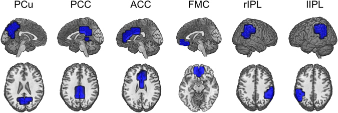
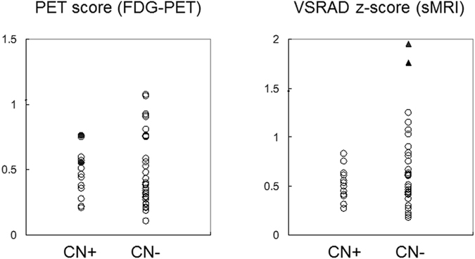
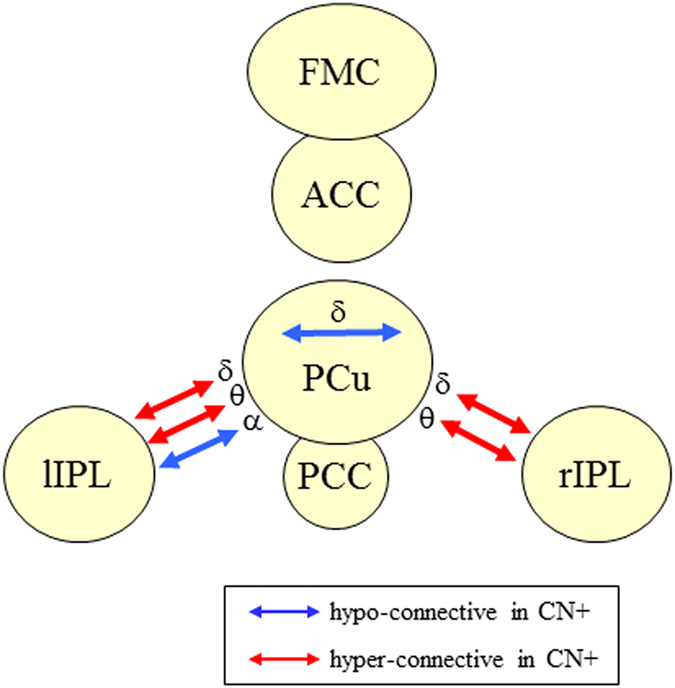
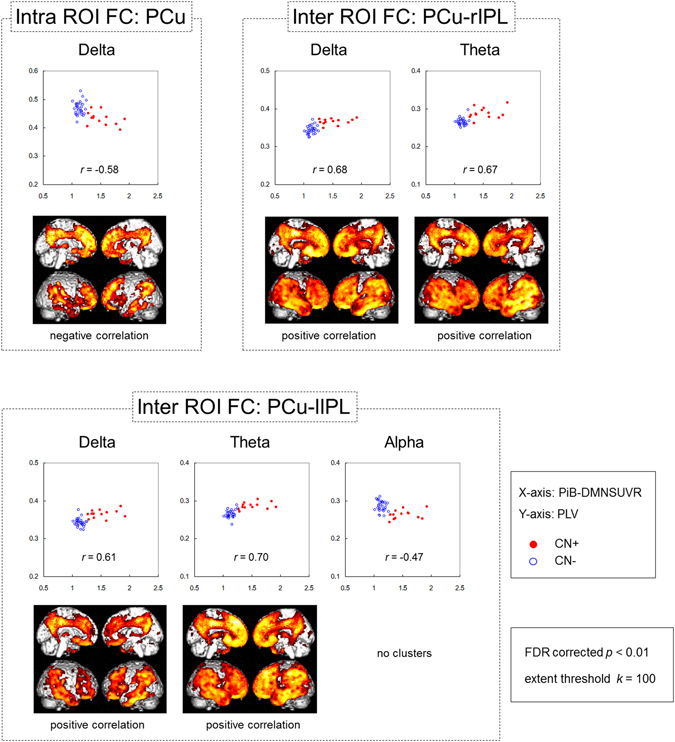
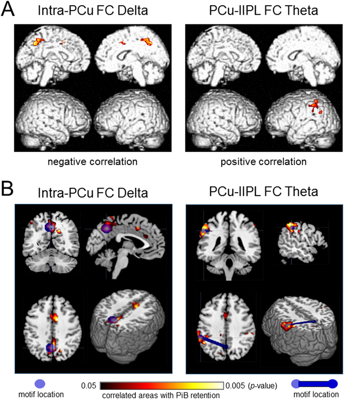
Amyloid-β (Aβ) deposition is known to starts decades before the onset of clinical symptoms of Alzheimer's disease (AD), however, the detailed pathophysiological processes underlying this preclinical period are not well understood. This study aimed to investigate functional network alterations in cognitively intact elderly individuals at risk for AD, and assessed the association between these network alterations and changes in Aβ deposition, glucose metabolism, and brain structure. Forty-five cognitively normal elderly subjects, who were classified into Aβ-positive (CN+) and Aβ-negative (CN-) groups using C-Pittsburgh compound B PET, underwent resting state magnetoencephalography measurements, F-fluorodeoxyglucose PET (FDG-PET) and structural MRI. Results demonstrated that in the CN+ group, functional connectivity (FC) within the precuneus was significantly decreased, whereas it was significantly enhanced between the precuneus and the bilateral inferior parietal lobules in the low-frequency bands (theta and delta). These changes were suggested to be associated with local cerebral Aβ deposition. Most of Aβ+ individuals in this study did not show any metabolic or anatomical changes, and there were no significant correlations between FC values and FDG-PET or MRI volumetry data. These results demonstrate that functional network alterations, which occur in association with Aβ deposition, are detectable using magnetoencephalography before metabolic and anatomical changes are seen.
淀粉样蛋白-β(Aβ)沉积已知在阿尔茨海默病(AD)临床症状出现前几十年就开始了,然而,这一临床前阶段的详细病理生理过程尚不清楚。本研究旨在研究认知正常的 AD 高危人群的功能网络改变,并评估这些网络改变与 Aβ沉积、葡萄糖代谢和脑结构变化之间的关系。45 名认知正常的老年受试者,根据 C-Pittsburgh 复合 B PET 分为 Aβ阳性(CN+)和 Aβ阴性(CN-)组,进行静息态脑磁图测量、F-氟脱氧葡萄糖正电子发射断层扫描(FDG-PET)和结构磁共振成像。结果表明,在 CN+组中,楔前叶内的功能连接(FC)显著降低,而在低频带(θ和δ)中,楔前叶与双侧顶下小叶之间的 FC 显著增强。这些变化被认为与局部脑 Aβ沉积有关。本研究中的大多数 Aβ+个体没有表现出任何代谢或解剖变化,FC 值与 FDG-PET 或 MRI 容积数据之间没有显著相关性。这些结果表明,在代谢和解剖变化出现之前,使用脑磁图可以检测到与 Aβ沉积相关的功能网络改变。