Choi Jiwoong, Hoffman Eric A, Lin Ching-Long, Milhem Mohammed M, Tessier Jean, Newell John D
Departments of Radiology, University of Iowa, Iowa City, Iowa, United States of America.
IIHR-Hydroscience & Engineering, University of Iowa, Iowa City, Iowa, United States of America.
PLoS One. 2017 Jul 27;12(7):e0179812. doi: 10.1371/journal.pone.0179812. eCollection 2017.
Extra-thoracic tumors send out pilot cells that attach to the pulmonary endothelium. We hypothesized that this could alter regional lung mechanics (tissue stiffening or accumulation of fluid and inflammatory cells) through interactions with host cells. We explored this with serial inspiratory computed tomography (CT) and image matching to assess regional changes in lung expansion.
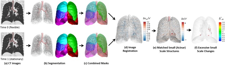
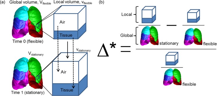
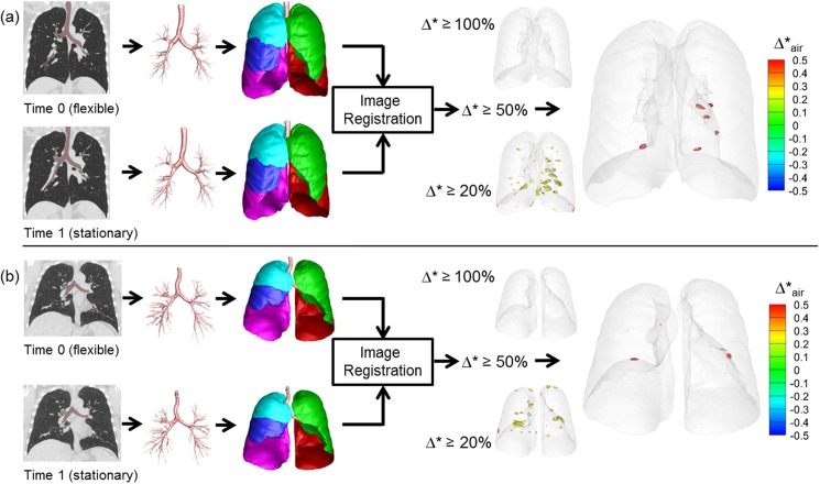
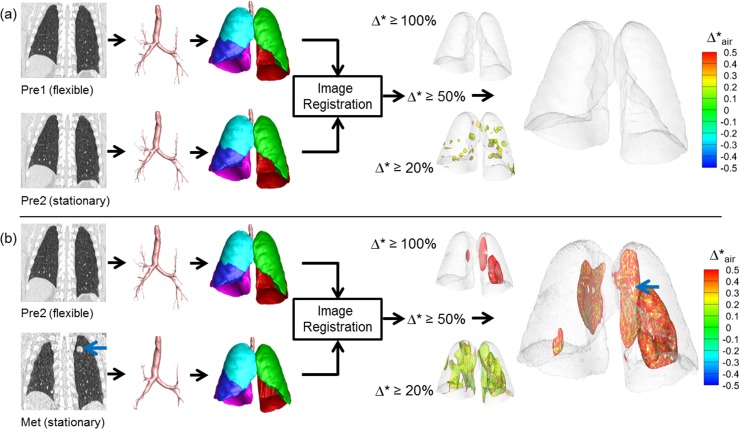
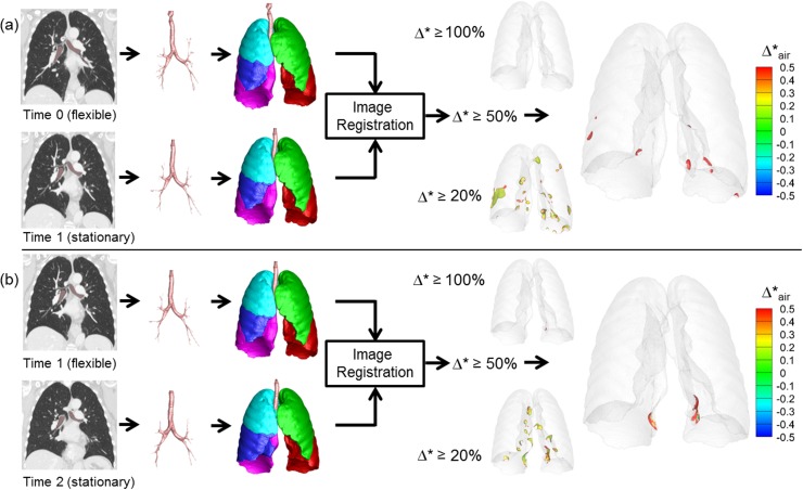
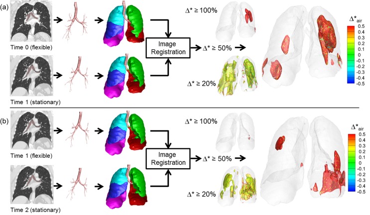
We retrospectively assessed 44 pairs of two serial CT scans on 21 sarcoma patients: 12 without lung metastases and 9 with lung metastases. For each subject, two or more serial inspiratory clinically-derived CT scans were retrospectively collected. Two research-derived control groups were included: 7 normal nonsmokers and 12 asymptomatic smokers with two inspiratory scans taken the same day or one year apart respectively. We performed image registration for local-to-local matching scans to baseline, and derived local expansion and density changes at an acinar scale. Welch two sample t test was used for comparison between groups. Statistical significance was determined with a p value < 0.05.
Lung regions of metastatic sarcoma patients (but not the normal control group) demonstrated an increased proportion of normalized lung expansion between the first and second CT. These hyper-expanded regions were associated with, but not limited to, visible metastatic lung lesions. Compared with the normal control group, the percent of increased normalized hyper-expanded lung in sarcoma subjects was significantly increased (p < 0.05). There was also evidence of increased lung "tissue" volume (non-air components) in the hyper-expanded regions of the cancer subjects relative to non-hyper-expanded regions. "Tissue" volume increase was present in the hyper-expanded regions of metastatic and non-metastatic sarcoma subjects. This putatively could represent regional inflammation related to the presence of tumor pilot cell-host related interactions.
This new quantitative CT (QCT) method for linking serial acquired inspiratory CT images may provide a diagnostic and prognostic means to objectively characterize regional responses in the lung following oncological treatment and monitoring for lung metastases.
胸外肿瘤会释放出附着于肺内皮的先导细胞。我们推测,这可能通过与宿主细胞的相互作用改变局部肺力学(组织硬化或液体及炎性细胞积聚)。我们通过连续吸气计算机断层扫描(CT)和图像匹配来探索这一现象,以评估肺扩张的局部变化。
我们回顾性评估了21例肉瘤患者的44对连续两次CT扫描:12例无肺转移患者和9例有肺转移患者。对于每个受试者,回顾性收集了两次或更多次连续吸气时临床获取的CT扫描。纳入了两个研究得出的对照组:7名正常非吸烟者和12名无症状吸烟者,分别在同一天或相隔一年进行了两次吸气扫描。我们对局部到局部的匹配扫描与基线进行图像配准,并在腺泡尺度上得出局部扩张和密度变化。采用韦尔奇两样本t检验进行组间比较。以p值<0.05确定统计学显著性。
转移性肉瘤患者的肺区域(而非正常对照组)在第一次和第二次CT之间显示出标准化肺扩张比例增加。这些过度扩张的区域与可见的转移性肺病变相关,但不限于这些病变。与正常对照组相比,肉瘤受试者中标准化过度扩张肺增加的百分比显著增加(p<0.05)。相对于非过度扩张区域,癌症受试者过度扩张区域的肺“组织”体积(非空气成分)也有增加的证据。转移性和非转移性肉瘤受试者的过度扩张区域均存在组织体积增加。这可能代表了与肿瘤先导细胞 - 宿主相关相互作用的存在有关的局部炎症。
这种用于关联连续获取的吸气CT图像中的新定量CT(QCT)方法,可能为客观表征肿瘤治疗后肺的局部反应以及监测肺转移提供一种诊断和预后手段。