Seyedmahmoud Rasoul, McGuire Jacob D, Wang Yong, Thiagarajan Ganesh, Walker Mary P
Department of Oral and Craniofacial Sciences, School of Dentistry, University of Missouri-Kansas City, MO, United States.
Department of Oral and Craniofacial Sciences, School of Dentistry, University of Missouri-Kansas City, MO, United States; Center of Excellence in Musculoskeletal and Dental Tissues, University of Missouri-Kansas City, MO, United States.
Dent Mater. 2017 Oct;33(10):1069-1074. doi: 10.1016/j.dental.2017.07.005. Epub 2017 Jul 24.
The aim of this paper is to determine the interrelationship between the microstructure - in terms of chemical composition and crystallinity - to the microhardness of coronal dentin.
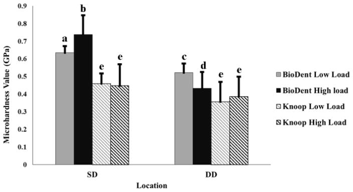
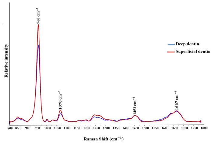
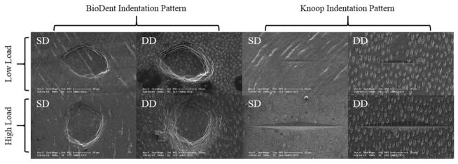
Dentin microhardness was tested by a novel reference point indenter and compared to the traditional Knoop hardness method. Micro-Raman spectroscopy was used to determine the chemical composition and crystallinity of dentin.
From the occlusal groove to the border of the coronal pulp chamber, dentin hardness decreased from superficial dentin (SD) to deep dentin (DD). Mineral/organic matrix ratios (phosphate/CH and phosphate/amide I) also decreased from SD to DD; however, this change was significant (P<0.05) in the phosphate/amide I ratio only. The phosphate/carbonate ratio decreased significantly by varying position from SD to DD. The degree of the crystallinity, as measured by the full width at half maximum (FWHM) of the peak at 960cm, decreased significantly going from superficial to deep dentin.
For the first time, the interrelationship between the microstructure and the mechanical properties of coronal dentin was determined by using the novel reference point indentation technique and micro-Raman spectroscopy. We hypothesize that the decrease in hardness from superficial to deep dentin can potentially be explained by decreased mineral content and increased carbonate content, which is also associated with decreased crystallinity. Collectively, there is a positive association between dentin hardness and mineral content and a negative association between dentin hardness and carbonate content.
本文旨在确定牙冠部牙本质在化学成分和结晶度方面的微观结构与显微硬度之间的相互关系。
采用一种新型参考点压头测试牙本质显微硬度,并与传统努氏硬度测试方法进行比较。利用显微拉曼光谱法测定牙本质的化学成分和结晶度。
从咬合沟到牙冠髓腔边界,牙本质硬度从表层牙本质(SD)到深层牙本质(DD)逐渐降低。矿物质/有机基质比率(磷酸盐/CH和磷酸盐/酰胺I)也从SD到DD逐渐降低;然而,只有磷酸盐/酰胺I比率的这种变化具有显著性(P<0.05)。磷酸盐/碳酸盐比率随位置从SD到DD显著降低。通过960cm处峰的半高宽(FWHM)测量的结晶度,从表层牙本质到深层牙本质显著降低。
首次利用新型参考点压痕技术和显微拉曼光谱法确定了牙冠部牙本质微观结构与力学性能之间的相互关系。我们推测,从表层牙本质到深层牙本质硬度的降低可能是由于矿物质含量降低和碳酸盐含量增加所致,这也与结晶度降低有关。总体而言,牙本质硬度与矿物质含量呈正相关,与碳酸盐含量呈负相关。