Department of Neuroscience, Karolinska Institutet, Retzius väg 8, SE-171 77, Stockholm, Sweden.
Department of Neurobiology, Care Sciences, and Society, Karolinska Institutet, Geriatrik-lab plan 5, SE-141 52, Huddinge, Sweden.
Neurotox Res. 2017 Nov;32(4):683-693. doi: 10.1007/s12640-017-9786-x. Epub 2017 Jul 29.
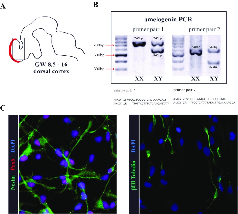
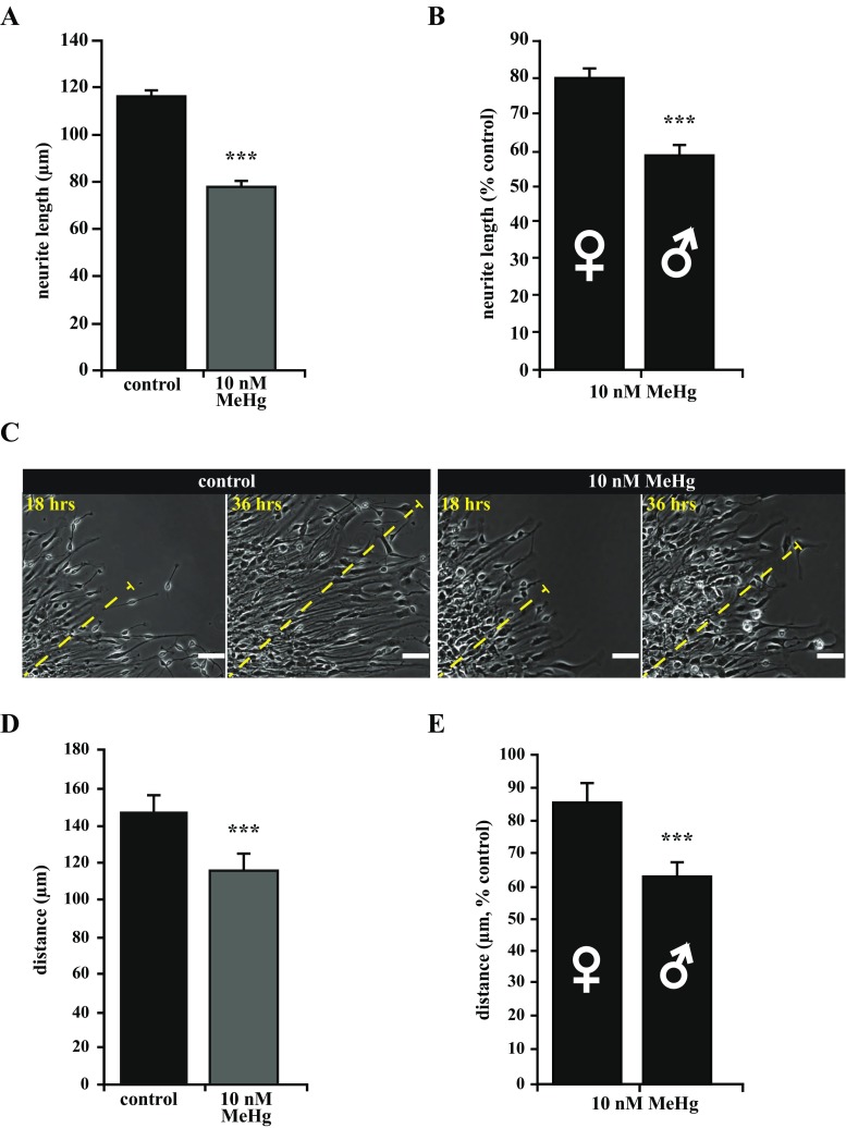
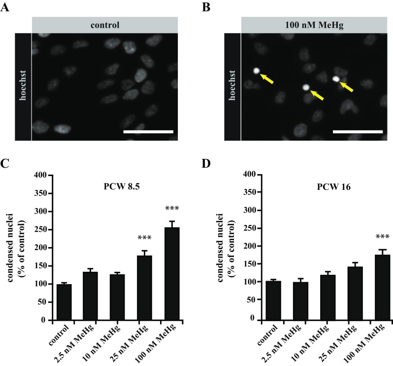
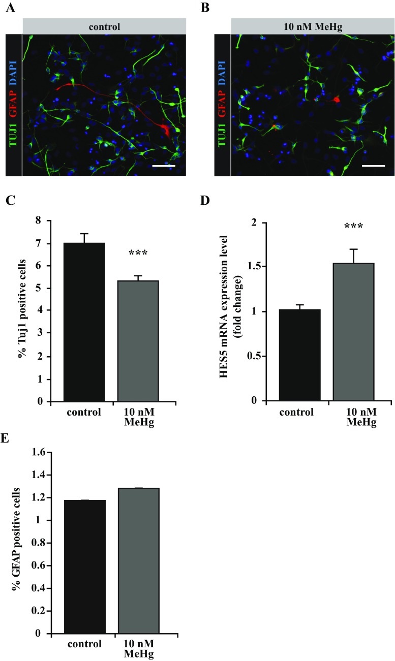
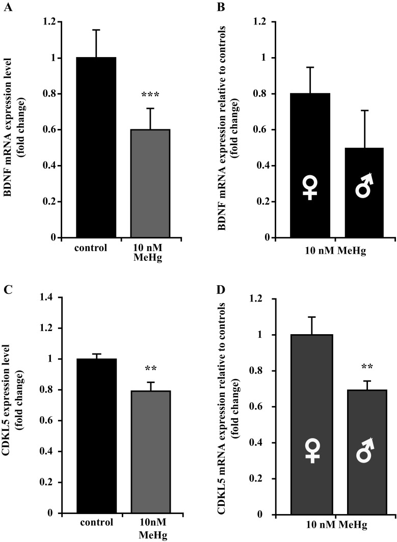
The developing nervous system is highly susceptible to methylmercury (MeHg), a widespread environmental neurotoxic contaminant. A wide range of morphological and functional outcomes have been described; however, there are still open questions regarding the mechanisms behind the developmental neurotoxic effects induced by low-level exposure. In the present study, we have examined the effects of nanomolar concentrations of MeHg on primary fetal human progenitor cells (hNPCs) with special focus on the role played by developmental stage and sex on the neurotoxic outcome. We found that neurospheres derived from earlier gestational time points exhibit higher susceptibility to MeHg, as they undergo apoptosis at a much lower dose (25 nM) as compared to neurospheres established from older fetuses (100 nM). At subapoptotic concentrations (10 nM), MeHg inhibited neuronal differentiation and maturation of hNPCs, as shown by a reduced number of Tuj1-positive cells and a visible reduction in neurite extension and cell migration, associated with a misregulation of Notch1 and BDNF signaling pathways. Interestingly, cells derived from male fetuses showed more severe alterations of neuronal morphology as compared to cells from females, indicating that the MeHg-induced impairment of neurite extension and cell migration is sex-dependent. Accordingly, the expression of the CDKL5 gene, a major factor regulating neurite outgrowth, was significantly more downregulated in male-derived cells. Altogether, gestational age and sex appear to be critical factors influencing in vitro hNPC sensitivity to low levels of MeHg.
发育中的神经系统对甲基汞(MeHg)高度敏感,甲基汞是一种广泛存在的环境神经毒性污染物。已经描述了广泛的形态和功能结果;然而,对于低水平暴露引起的发育神经毒性作用的机制仍存在一些悬而未决的问题。在本研究中,我们研究了纳摩尔浓度的 MeHg 对原代人胎儿祖细胞(hNPC)的影响,特别关注发育阶段和性别对神经毒性结果的影响。我们发现,来自更早妊娠时间点的神经球对 MeHg 的敏感性更高,因为它们在低得多的剂量(25 nM)下发生凋亡,而来自较晚胎儿的神经球在 100 nM 时才发生凋亡。在亚凋亡浓度(10 nM)下,MeHg 抑制 hNPC 的神经元分化和成熟,这表现为 Tuj1 阳性细胞数量减少,以及神经突延伸和细胞迁移的明显减少,与 Notch1 和 BDNF 信号通路的失调有关。有趣的是,与来自女性的细胞相比,来自男性胎儿的细胞显示出更严重的神经元形态改变,表明 MeHg 诱导的神经突延伸和细胞迁移损伤是性别依赖性的。相应地,CDKL5 基因的表达,一种调节神经突生长的主要因素,在男性来源的细胞中明显下调更多。总之,胎龄和性别似乎是影响 hNPC 对低水平 MeHg 敏感性的关键因素。