Jung Christian, Grün Katja, Betge Stefan, Pernow John, Kelm Malte, Muessig Johanna, Masyuk Maryna, Kuethe Friedhelm, Ndongson-Dongmo Bernadin, Bauer Reinhard, Lauten Alexander, Schulze P Christian, Berndt Alexander, Franz Marcus
Department of Internal Medicine, Division of Cardiology, Pulmonology and Vascular Medicine, University Hospital Düsseldorf, Heinrich-Heine-University, Düsseldorf 40225, Germany.
Department of Internal Medicine I, Jena University Hospital, Jena 07747, Germany.
Int J Mol Sci. 2017 Jul 25;18(8):1609. doi: 10.3390/ijms18081609.
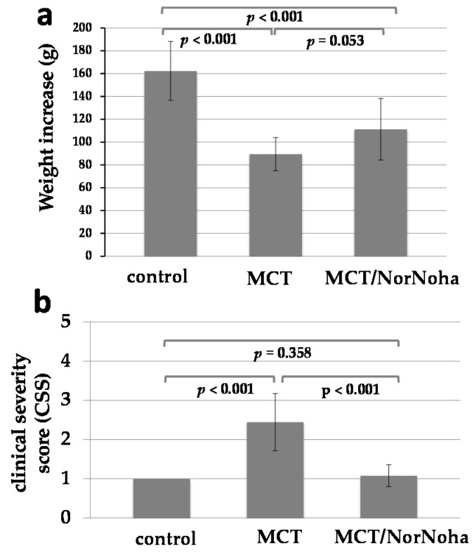
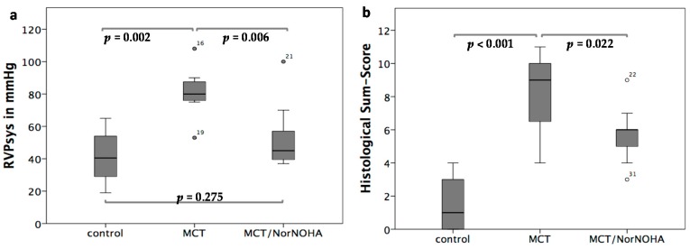
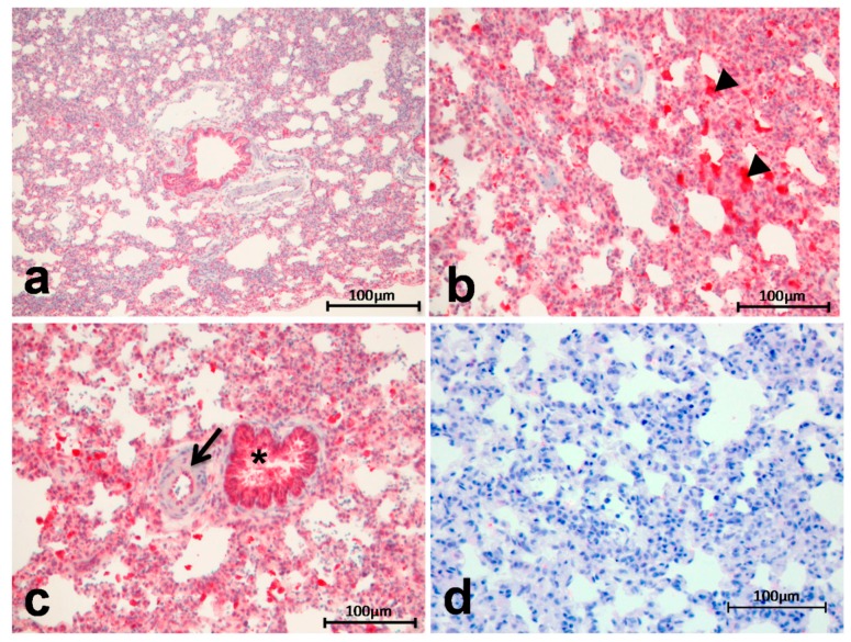
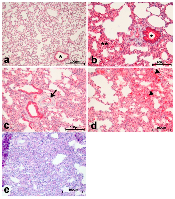
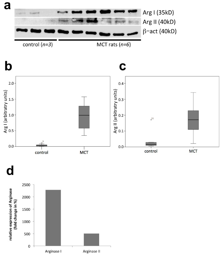
Pulmonary hypertension (PH) is a heterogeneous disorder associated with a poor prognosis. Thus, the development of novel treatment strategies is of great interest. The enzyme arginase (Arg) is emerging as important player in PH development. The aim of the current study was to determine the expression of ArgI and ArgII as well as the effects of Arg inhibition in a rat model of PH. PH was induced in 35 Sprague-Dawley rats by monocrotaline (MCT, 60 mg/kg as single-dose). There were three experimental groups: sham-treated controls (control group, = 11), MCT-induced PH (MCT group, = 11) and MCT-induced PH treated with the Arg inhibitor Nω-hydroxy-nor-l-arginine (nor-NOHA; MCT/NorNoha group, = 13). ArgI and ArgII expression was determined by immunohistochemistry and Western blot. Right ventricular systolic pressure (RVPsys) was measured and lung tissue remodeling was determined. Induction of PH resulted in an increase in RVPsys (81 ± 16 mmHg) compared to the control group (41 ± 15 mmHg, = 0.002) accompanied by a significant elevation of histological sum-score (8.2 ± 2.4 in the MCT compared to 1.6 ± 1.6 in the control group, < 0.001). Both, ArgI and ArgII were relevantly expressed in lung tissue and there was a significant increase in the MCT compared to the control group ( < 0.01). Arg inhibition resulted in a significant reduction of RVPsys to 52 ± 19 mmHg ( = 0.006) and histological sum-score to 5.8 ± 1.4 compared to the MCT group ( = 0.022). PH leads to increased expression of Arg. Arg inhibition leads to reduction of RVPsys and diminished lung tissue remodeling and therefore represents a potential treatment strategy in PH.
肺动脉高压(PH)是一种预后不良的异质性疾病。因此,新型治疗策略的开发备受关注。精氨酸酶(Arg)作为PH发展过程中的重要参与者正逐渐崭露头角。本研究的目的是确定精氨酸酶I(ArgI)和精氨酸酶II(ArgII)的表达以及Arg抑制在PH大鼠模型中的作用。通过给予35只Sprague-Dawley大鼠一次性腹腔注射野百合碱(MCT,60mg/kg)诱导PH。实验分为三组:假手术对照组(对照组,n = 11)、MCT诱导的PH组(MCT组,n = 11)和用Arg抑制剂Nω-羟基-L-精氨酸(nor-NOHA)治疗的MCT诱导的PH组(MCT/NorNoha组,n = 13)。通过免疫组织化学和蛋白质印迹法测定ArgI和ArgII的表达。测量右心室收缩压(RVPsys)并确定肺组织重塑情况。与对照组(41±15mmHg,P = 0.002)相比,PH诱导导致RVPsys升高(81±16mmHg),同时组织学总分显著升高(MCT组为8.2±2.4,对照组为1.6±1.6,P < 0.001)。ArgI和ArgII在肺组织中均有相关表达,与对照组相比,MCT组显著增加(P < 0.01)。与MCT组相比,Arg抑制导致RVPsys显著降低至52±19mmHg(P = 0.006),组织学总分降低至5.8±1.4(P = 0.022)。PH导致Arg表达增加。Arg抑制导致RVPsys降低和肺组织重塑减轻,因此是PH的一种潜在治疗策略。