Division of Cardiology, Department of Internal Medicine, Tongji Hospital, Tongji Medical College of Huazhong University of Science and Technology, Hubei Key Laboratory of Genetics and Molecular Mechanisms of Cardiological Disorders, Wuhan, 430030, China.
Sci Rep. 2017 Jul 31;7(1):6926. doi: 10.1038/s41598-017-07512-1.
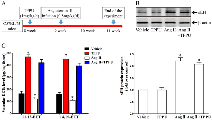
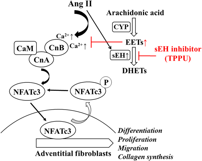
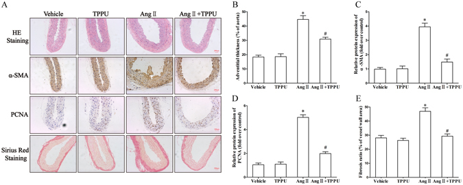
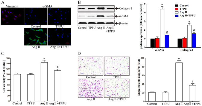
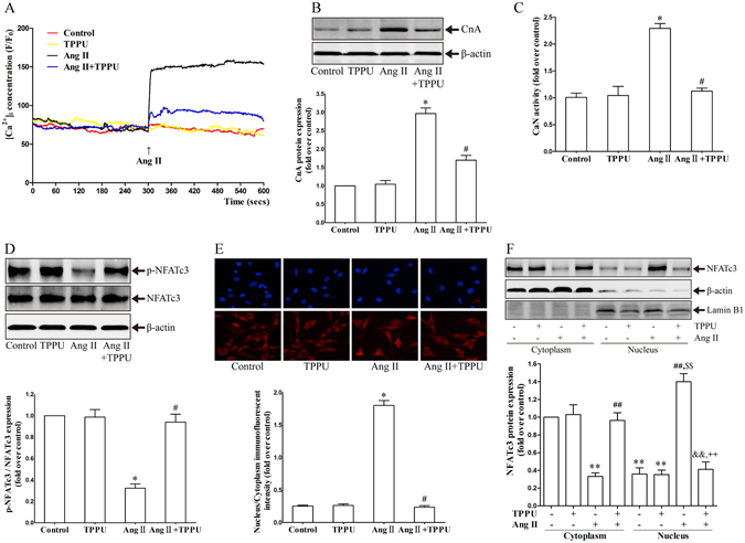
Epoxyeicosatrienoic acids (EETs), the metabolites of cytochrome P450 epoxygenases derived from arachidonic acid, exert important biological activities in maintaining cardiovascular homeostasis. Soluble epoxide hydrolase (sEH) hydrolyzes EETs to less biologically active dihydroxyeicosatrienoic acids. However, the effects of sEH inhibition on adventitial remodeling remain inconclusive. In this study, the adventitial remodeling model was established by continuous Ang II infusion for 2 weeks in C57BL/6 J mice, before which sEH inhibitor 1-trifluoromethoxyphenyl-3-(1-propionylpiperidin-4-yl) urea (TPPU) was administered by gavage. Adventitial remodeling was evaluated by histological analysis, western blot, immunofluorescent staining, calcium imaging, CCK-8 and transwell assay. Results showed that Ang II infusion significantly induced vessel wall thickening, collagen deposition, and overexpression of α-SMA and PCNA in aortic adventitia, respectively. Interestingly, these injuries were attenuated by TPPU administration. Additionally, TPPU pretreatment overtly prevented Ang II-induced primary adventitial fibroblasts activation, characterized by differentiation, proliferation, migration, and collagen synthesis via Ca-calcineurin/NFATc3 signaling pathway in vitro. In summary, our results suggest that inhibition of sEH could be considered as a novel therapeutic strategy to treat adventitial remodeling related disorders.
环氧二十碳三烯酸(EETs)是花生四烯酸衍生的细胞色素 P450 加氧酶的代谢产物,在维持心血管稳态方面发挥着重要的生物学作用。可溶性环氧化物水解酶(sEH)将 EETs 水解为生物活性较低的二羟二十碳三烯酸。然而,sEH 抑制对外膜重塑的影响仍存在争议。在本研究中,通过连续 Ang II 输注 2 周在 C57BL/6J 小鼠中建立外膜重塑模型,在此之前通过灌胃给予 sEH 抑制剂 1-三氟甲氧基苯基-3-(1-丙酰基哌啶-4-基)脲(TPPU)。通过组织学分析、western blot、免疫荧光染色、钙成像、CCK-8 和 Transwell 测定评估外膜重塑。结果表明,Ang II 输注显著诱导血管壁增厚、胶原沉积以及主动脉外膜中 α-SMA 和 PCNA 的过度表达。有趣的是,这些损伤通过 TPPU 给药得到了缓解。此外,TPPU 预处理明显阻止了 Ang II 诱导的原代外膜成纤维细胞激活,其特征是体外分化、增殖、迁移和胶原合成,这是通过 Ca-钙调神经磷酸酶/NFATc3 信号通路介导的。综上所述,我们的结果表明,抑制 sEH 可能被认为是治疗外膜重塑相关疾病的一种新的治疗策略。