Pratesi Giovanni, Caporali Stefano, Loglio Francesca, Giuli Gabriele, Dziková Lenka, Skála Roman
Museo di Storia Naturale, Università di Firenze, Via G. La Pira 4, 50121 Firenze, Italy.
Department of Chemistry, Università di Firenze, Via della Lastruccia 3, 50019 Sesto Fiorentino, Italy.
Materials (Basel). 2014 Apr 24;7(4):3319-3336. doi: 10.3390/ma7043319.
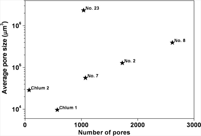
X-ray micro-computer aided tomography (μ-CT), together with optical microscopy and imaging, have been applied to the study of six moldavite samples. These techniques enabled a complete characterization to be made of the textural features of both Muong Nong-type and common splashform moldavites. A detailed study of the size and distribution of pores or bubbles confirmed the marked variability in pore size among the samples, as well as within each sample, and indicated in the Muong Nong-type moldavites the presence of at least two deformation stages which occurred before and after pore formation.
X射线微计算机断层扫描(μ-CT)与光学显微镜和成像技术一起,已被应用于对六个捷克玻陨石样本的研究。这些技术能够对Muong Nong型和普通溅落型捷克玻陨石的结构特征进行全面表征。对孔隙或气泡的大小和分布进行的详细研究证实了样本之间以及每个样本内部孔隙大小的显著差异,并表明在Muong Nong型捷克玻陨石中至少存在两个在孔隙形成之前和之后发生的变形阶段。