Thaler Christian, Schneider Tanja, Sedlacik Jan, Kutzner Daniel, Stellmann Jan-Patrick, Heesen Christoph, Fiehler Jens, Siemonsen Susanne
Department of Diagnostic and Interventional Neuroradiology, University Medical Centre Hamburg-Eppendorf, Hamburg, Germany.
Department of Neurology, University Medical Centre Hamburg-Eppendorf, Hamburg, Germany.
PLoS One. 2017 Aug 10;12(8):e0183099. doi: 10.1371/journal.pone.0183099. eCollection 2017.
In multiple sclerosis (MS) the sensitivity for detection of contrast enhancing lesions (CEL) in T1-weighted scans is essential for diagnostics and therapy decisions. The purpose of our study was to evaluate the sensitivity of T1w MPRAGE scans in comparison to T1w dark blood technique (T1-DB) for CEL in MS.
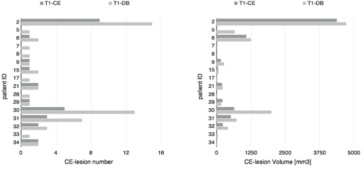
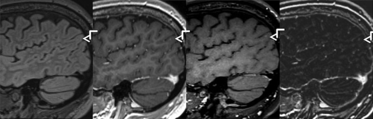
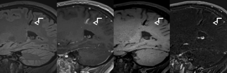
3T MR imaging was performed in 37 MS patients, including T2-weighted imaging, T1w MPRAGE before and after gadolinium injection (unenhanced-T1 and T1-CE) and T1-DB imaging. After gadolinium application, the T1-DB scan was performed prior to T1-CE. From unenhanced-T1 and T1-CE scans, subtraction images (T1-SUB) were calculated. The number of CEL was determined separately on T1-CE and T1-DB by two raters independently. Lesions only detected on T1-DB scans then were verified on T1-SUB. Only lesions detected by both raters were included in further analysis.
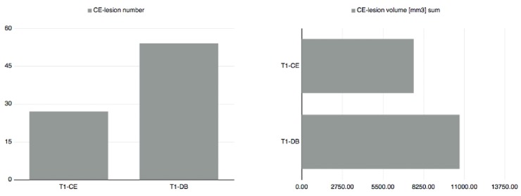
In 16 patients, at least one CEL was detected by both rater, either on T1-CE or T1-DB. All lesions that were detected on T1-CE were also detected on T1-DB images. The total number of contrast enhancing lesions detected on T1-DB images (n = 54) by both raters was significantly higher than the corresponding number of lesions identified on T1-CE (n = 27) (p = 0.01); all of these lesions could be verified on SUB images. In 21 patients, no CEL was detected in any of the sequences.
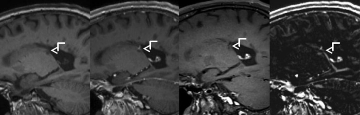
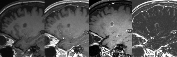
The application of T1-DB technique increases the sensitivity for CEL in MS, especially for those lesions that show only subtle increase in intensity after Gadolinium application but remain hypo- or iso-intense to surrounding tissue.
在多发性硬化症(MS)中,T1加权扫描中检测对比增强病灶(CEL)的敏感性对于诊断和治疗决策至关重要。我们研究的目的是评估T1加权MPRAGE扫描与T1加权黑血技术(T1-DB)相比对MS中CEL的敏感性。
对37例MS患者进行3T磁共振成像,包括T2加权成像、钆注射前后的T1加权MPRAGE(未增强T1和T1-CE)以及T1-DB成像。应用钆后,在T1-CE之前进行T1-DB扫描。从未增强T1和T1-CE扫描中计算减法图像(T1-SUB)。两名评估者分别在T1-CE和T1-DB上确定CEL的数量。然后在T1-SUB上验证仅在T1-DB扫描中检测到的病灶。仅两名评估者均检测到的病灶纳入进一步分析。
在16例患者中,两名评估者在T1-CE或T1-DB上均检测到至少一个CEL。在T1-CE上检测到的所有病灶在T1-DB图像上也被检测到。两名评估者在T1-DB图像上检测到的对比增强病灶总数(n = 54)显著高于在T1-CE上识别的相应病灶数(n = 27)(p = 0.01);所有这些病灶均可在SUB图像上得到验证。在21例患者中,任何序列均未检测到CEL。
T1-DB技术的应用提高了MS中CEL的敏感性,特别是对于那些在应用钆后强度仅轻微增加但对周围组织仍呈低或等强度的病灶。