A. A. Martinos Center for Biomedical Imaging, Department of Radiology, Massachusetts General Hospital and Harvard Medical School, 149 Thirteenth St., Suite 2301, Charlestown, MA, 02129, USA.
Institute of Organic Chemistry and Biochemistry of the Czech Academy of Sciences, Flemingovo nam. 2, 16610, Prague 6, Czech Republic.
Sci Rep. 2017 Aug 14;7(1):8114. doi: 10.1038/s41598-017-08838-6.
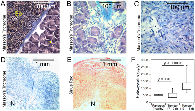
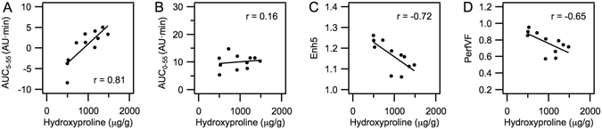
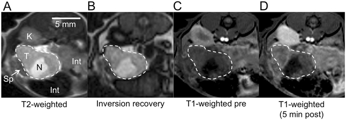
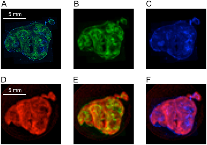
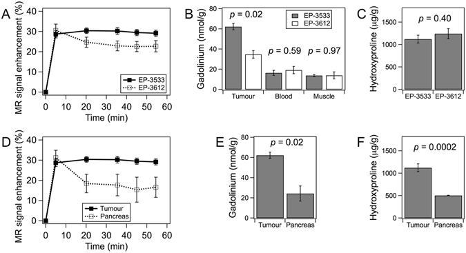
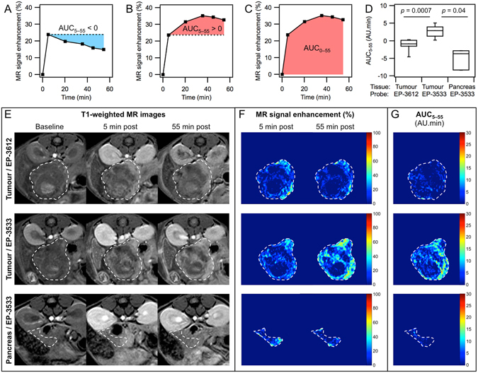
Fibrosis with excessive amounts of type I collagen is a hallmark of many solid tumours, and fibrosis is a promising target in cancer therapy, but tools for its non-invasive quantification are missing. Here we used magnetic resonance imaging with a gadolinium-based probe targeted to type I collagen (EP-3533) to image and quantify fibrosis in pancreatic ductal adenocarcinoma. An orthotopic syngeneic mouse model resulted in tumours with 2.3-fold higher collagen level compared to healthy pancreas. Animals were scanned at 4.7 T before, during and up to 60 min after i.v. injection of EP-3533, or of its non-binding isomer EP-3612. Ex-vivo quantification of gadolinium showed significantly higher uptake of EP-3533 compared to EP-3612 in tumours, but not in surrounding tissue (blood, muscle). Uptake of EP-3533 visualized in T1-weighted MRI correlated well with spatial distribution of collagen determined by second harmonic generation imaging. Differences in the tumour pharmacokinetic profiles of EP-3533 and EP-3612 were utilized to distinguish specific binding to tumour collagen from non-specific uptake. A model-free pharmacokinetic measurement based on area under the curve was identified as a robust imaging biomarker of fibrosis. Collagen-targeted molecular MRI with EP-3533 represents a new tool for non-invasive visualization and quantification of fibrosis in tumour tissue.
纤维化为多种实体瘤的显著特征,且纤维化是癌症治疗中极具潜力的靶点,但目前尚缺乏针对其的非侵入性定量方法。本研究使用针对 I 型胶原蛋白的镧系元素探针(EP-3533)进行磁共振成像,以对胰腺导管腺癌中的纤维化进行成像和定量分析。在该同基因原位小鼠模型中,肿瘤的胶原蛋白水平较健康胰腺高 2.3 倍。在 4.7T 下对动物进行扫描,分别在静脉注射 EP-3533 及其非结合异构体 EP-3612 之前、期间以及之后的 60 分钟内进行扫描。对体外的钆定量分析显示,EP-3533 在肿瘤中的摄取明显高于 EP-3612,但在周围组织(血液、肌肉)中无差异。T1 加权 MRI 中观察到的 EP-3533 摄取与由二次谐波产生成像确定的胶原蛋白的空间分布密切相关。EP-3533 和 EP-3612 在肿瘤中的药代动力学特征差异被用于区分其与肿瘤胶原蛋白的特异性结合与非特异性摄取。基于曲线下面积的无模型药代动力学测量被确定为纤维化的稳健成像生物标志物。利用 EP-3533 进行的靶向胶原蛋白的分子 MRI 为肿瘤组织中的纤维化进行非侵入性可视化和定量提供了一种新方法。