Department of Perioperative Medicine, Clinical Center, National Institutes of Health, Bethesda, MD 20892.
Biotherapy Section, Laboratory of Molecular Biology, Center for Cancer Research, National Cancer Institute, Bethesda, MD 20892.
Mol Pain. 2017 Jan-Dec;13:1744806917727657. doi: 10.1177/1744806917727657.
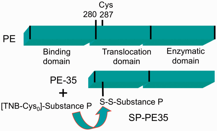
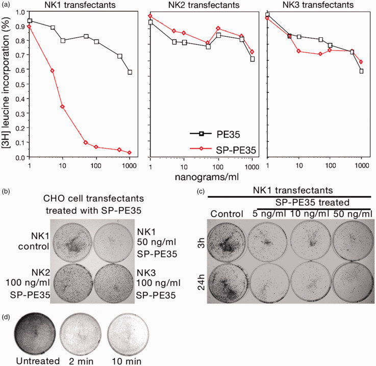
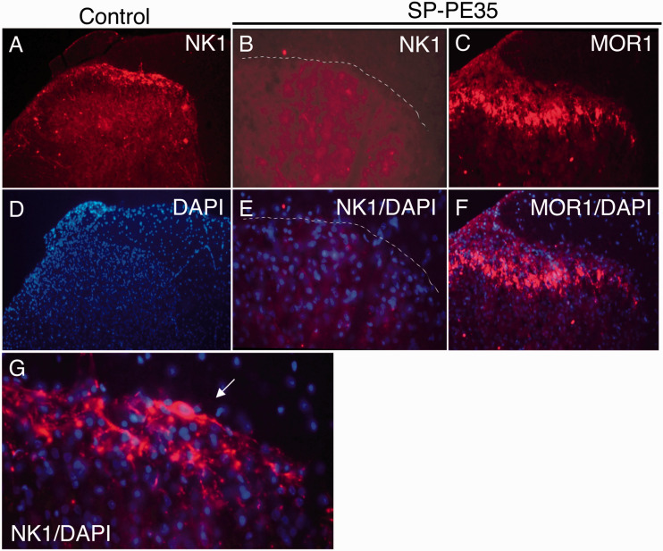
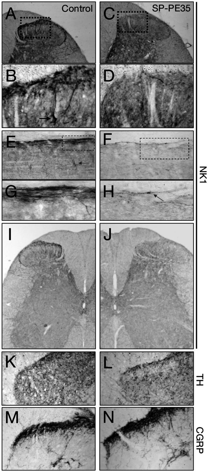
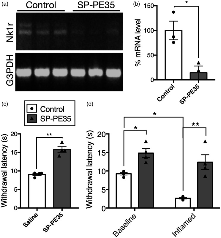
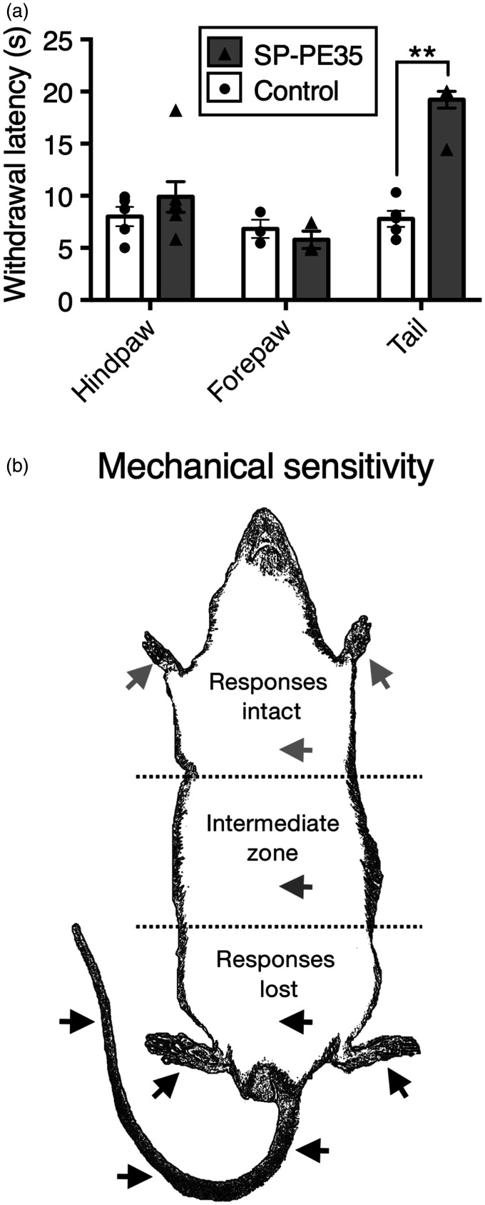
Cell deletion approaches to pain directed at either the primary nociceptive afferents or second-order neurons are highly effective analgesic manipulations. Second-order spinal neurons expressing the neurokinin 1 (NK1) receptor are required for the perception of many types of pain. To delete NK1+ neurons for the purpose of pain control, we generated a toxin–peptide conjugate using DTNB-derivatized (Cys0) substance P (SP) and a N-terminally truncated Pseudomonas exotoxin (PE35) that retains the endosome-release and ADP-ribosylation enzymatic domains but with only one free sulfhydryl side chain for conjugation. This allowed generation of a one-to-one product linked by a disulfide bond (SP-PE35). In vitro, Chinese hamster ovary cells stably transfected with the NK1 receptor exhibited specific cytotoxicity when exposed to SP-PE35 (IC50 = 5 × 10⁻¹¹ M), whereas the conjugate was nontoxic to NK2 and NK3 receptor-bearing cell lines. In vivo studies showed that, after infusion into the spinal subarachnoid space, the toxin was extremely effective in deleting NK1 receptor-expressing cells from the dorsal horn of the spinal cord. The specific cell deletion robustly attenuated thermal and mechanical pain sensations and inflammatory hyperalgesia but did not affect motoric capabilities. NK1 receptor cell deletion and antinociception occurred without obvious lesion of non–receptor-expressing cells or apparent reorganization of primary afferent innervation. These data demonstrate the extraordinary selectivity and broad-spectrum antinociceptive efficacy of this ligand-directed protein therapeutic acting via receptor-mediated endocytosis. The loss of multiple pain modalities including heat and mechanical pinch, transduced by different populations of primary afferents, shows that spinal NK1 receptor-expressing neurons are critical points of convergence in the nociceptive transmission circuit. We further suggest that therapeutic end points can be effectively and safely achieved when SP-PE35 is locally infused, thereby producing a regionally defined analgesia.
针对初级伤害感受传入神经或二级神经元的细胞删除方法是非常有效的镇痛操作。表达神经激肽 1 (NK1)受体的二级脊髓神经元是许多类型疼痛感知所必需的。为了删除 NK1+神经元以控制疼痛,我们使用 DTNB 衍生的 (Cys0) 物质 P (SP) 和截短的假单胞菌外毒素 (PE35) 生成了一种毒素-肽缀合物,该缀合物保留了内体释放和 ADP-核糖基化酶结构域,但只有一个游离的巯基侧链用于缀合。这允许生成通过二硫键连接的一对一产物 (SP-PE35)。在体外,稳定转染 NK1 受体的中国仓鼠卵巢细胞在暴露于 SP-PE35 时表现出特异性细胞毒性 (IC50 = 5 × 10⁻¹¹ M),而该缀合物对携带 NK2 和 NK3 受体的细胞系没有毒性。体内研究表明,在蛛网膜下腔输注后,毒素非常有效地从脊髓背角中删除表达 NK1 受体的细胞。特异性细胞删除强烈减弱了热和机械痛觉以及炎症性痛觉过敏,但不影响运动能力。NK1 受体细胞缺失和镇痛作用发生而没有明显的非受体表达细胞的损伤或初级传入神经支配的明显重组。这些数据表明,这种配体导向的蛋白质治疗通过受体介导的内吞作用具有非凡的选择性和广谱的镇痛疗效。包括热和机械夹捏在内的多种疼痛方式的丧失,由不同的初级传入神经元群传递,表明脊髓 NK1 受体表达神经元是伤害性传递回路中的关键点。我们进一步表明,当 SP-PE35 局部输注时,可以有效地和安全地达到治疗终点,从而产生区域定义的镇痛作用。