Department of Mountain and Sleep Medicine and Pulmonary Hypertension, National Center of Cardiology and Internal Medicine, Bishkek, Kyrgyzstan.
Kyrgyz-Indian Mountain Biomedical Research Center, Bishkek, Kyrgyzstan.
Can Respir J. 2017;2017:1587865. doi: 10.1155/2017/1587865. Epub 2017 Jul 26.
Recent studies have reported that obstructive sleep apnea (OSA) patients present alterations in right ventricular (RV) structure and function. However, large randomized controlled trials evaluating the impact of OSA on the right ventricle are lacking.
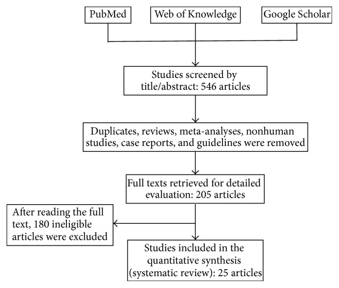
A comprehensive electronic database (PubMed, Web of Science, and Google Scholar) and reference search up to October 30, 2016, was performed. A systematic review and meta-analysis were performed to assess RV structure and function in OSA patients based on conventional echocardiography and tissue Doppler imaging.
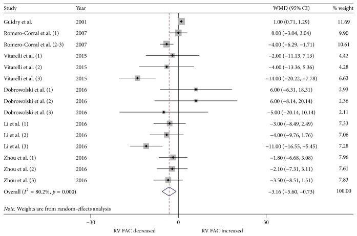
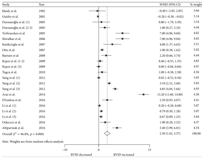
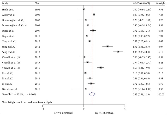
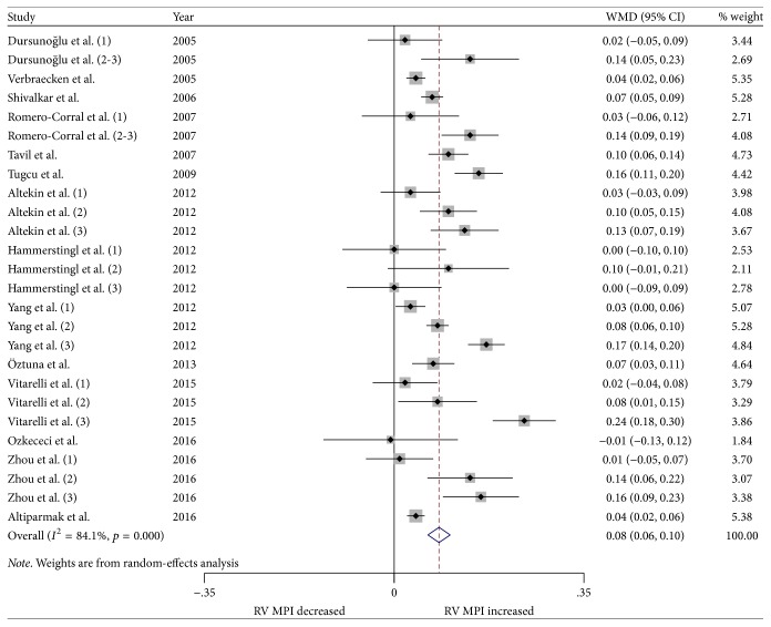
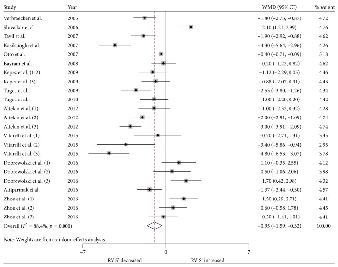
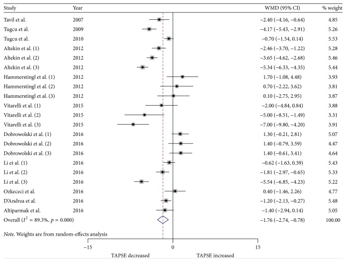
Twenty-five studies with 1,503 OSA patients and 796 controls were included in this study. OSA patients exhibited an increase in RV internal diameter (weighted mean difference (WMD) (95% confidence intervals (CIs)) 2.49 (1.62 to 3.37); = 0.000) and RV wall thickness (WMD (95% CIs) 0.82 (0.51 to 1.13); = 0.000). Furthermore, OSA patients had a significantly elevated RV myocardial performance index (WMD (95% CI) 0.08 (0.06 to 0.10); = 0.000), decreased RV S' (WMD (95% CI) -0.95 (-1.59 to -0.32); = 0.003), tricuspid annular plane systolic excursion (WMD (95% CI) -1.76 (-2.73 to -0.78); = 0.000), and RV fractional area change (WMD (95% CI) -3.16 (-5.60 to -0.73); = 0.011).
OSA patients display RV dilatation, increased wall thickening, and altered RV function.
最近的研究报告称,阻塞性睡眠呼吸暂停(OSA)患者的右心室(RV)结构和功能发生改变。然而,缺乏评估 OSA 对右心室影响的大型随机对照试验。
对截至 2016 年 10 月 30 日的综合电子数据库(PubMed、Web of Science 和 Google Scholar)和参考文献进行了检索。基于常规超声心动图和组织多普勒成像,进行了系统评价和荟萃分析,以评估 OSA 患者的 RV 结构和功能。
共纳入 25 项研究,其中 1503 例 OSA 患者和 796 例对照者。OSA 患者的 RV 内径增加(加权均数差(WMD)(95%置信区间(CI))2.49(1.62 至 3.37); = 0.000)和 RV 壁厚度增加(WMD(95%CI)0.82(0.51 至 1.13); = 0.000)。此外,OSA 患者的 RV 心肌做功指数明显升高(WMD(95%CI)0.08(0.06 至 0.10); = 0.000),RV S'降低(WMD(95%CI)-0.95(-1.59 至 -0.32); = 0.003),三尖瓣环平面收缩位移(WMD(95%CI)-1.76(-2.73 至 -0.78); = 0.000)和 RV 节段面积变化率(WMD(95%CI)-3.16(-5.60 至 -0.73); = 0.011)。
OSA 患者表现出 RV 扩张、壁增厚和 RV 功能改变。