Department of Medicine, Veterans Affairs Medical Center, Denver, Colorado.
Department of Medicine, University of Colorado Anschutz Medical Campus, Aurora, Colorado.
Cancer Immunol Res. 2017 Sep;5(9):767-777. doi: 10.1158/2326-6066.CIR-16-0365. Epub 2017 Aug 17.
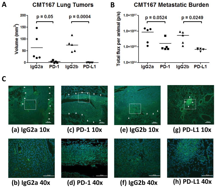
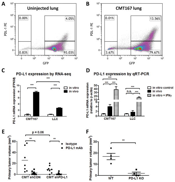
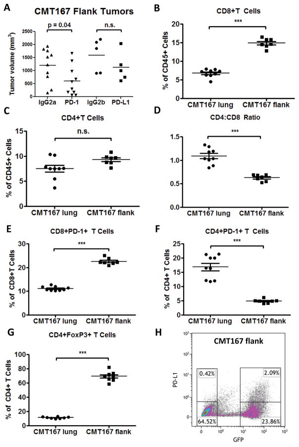
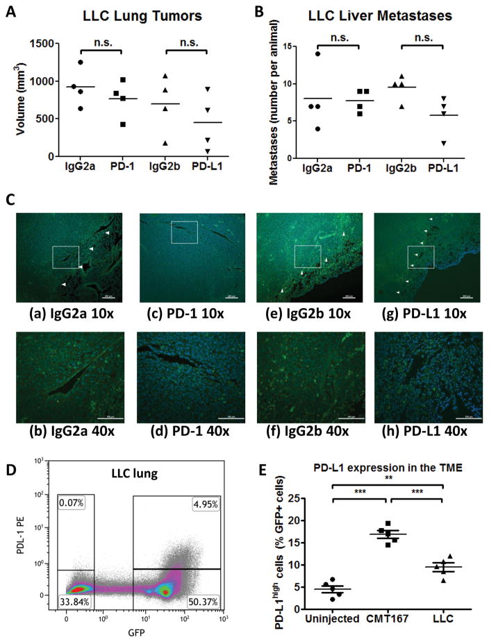
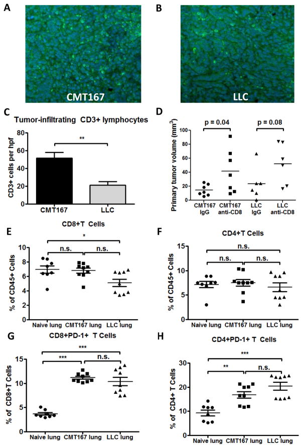
Immune checkpoint inhibitors targeting the interaction between programmed cell death-1 (PD-1) and its ligand PD-L1 induce tumor regression in a subset of non-small cell lung cancer patients. However, clinical response rates are less than 25%. Evaluation of combinations of immunotherapy with existing therapies requires appropriate preclinical animal models. In this study, murine lung cancer cells (CMT167 and LLC) were implanted either orthotopically in the lung or subcutaneously in syngeneic mice, and response to anti-PD-1/PD-L1 therapy was determined. Anti-PD-1/PD-L1 therapy inhibited CMT167 orthotopic lung tumors by 95%. The same treatments inhibited CMT167 subcutaneous tumors by only 30% and LLC orthotopic lung tumors by 35%. CMT167 subcutaneous tumors had more Foxp3 CD4 T cells and fewer PD-1 CD4 T cells compared with CMT167 orthotopic tumors. Flow cytometric analysis also demonstrated increased abundance of PD-L1 cells in the tumor microenvironment in CMT167 tumor-bearing lungs compared with CMT167 subcutaneous tumors or LLC tumor-bearing lungs. Silencing PD-L1 expression in CMT167 cells resulted in smaller orthotopic tumors that remained sensitive to anti-PD-L1 therapy, whereas implantation of CMT167 cells into PD-L1 mice blocked orthotopic tumor growth, indicating a role for PD-L1 in both the cancer cell and the microenvironment. These findings indicate that the response of cancer cells to immunotherapy will be determined by both intrinsic properties of the cancer cells and specific interactions with the microenvironment. Experimental models that accurately recapitulate the lung tumor microenvironment are useful for evaluation of immunotherapeutic agents. .
免疫检查点抑制剂靶向程序性细胞死亡-1(PD-1)与其配体 PD-L1 之间的相互作用,可诱导部分非小细胞肺癌患者的肿瘤消退。然而,临床反应率低于 25%。评估免疫疗法与现有疗法的联合应用需要适当的临床前动物模型。在这项研究中,将鼠肺癌细胞(CMT167 和 LLC)分别原位植入肺或同基因小鼠皮下,并确定对抗 PD-1/PD-L1 治疗的反应。抗 PD-1/PD-L1 治疗抑制 CMT167 原位肺肿瘤达 95%。相同的治疗方法仅抑制 CMT167 皮下肿瘤 30%和 LLC 原位肺肿瘤 35%。与 CMT167 原位肿瘤相比,CMT167 皮下肿瘤的 Foxp3 CD4 T 细胞更多,PD-1 CD4 T 细胞更少。流式细胞术分析还表明,与 CMT167 皮下肿瘤或 LLC 荷瘤肺相比,CMT167 荷瘤肺中的肿瘤微环境中 PD-L1 细胞的丰度增加。CMT167 细胞中 PD-L1 表达的沉默导致原位肿瘤变小,仍然对抗 PD-L1 治疗敏感,而将 CMT167 细胞植入 PD-L1 小鼠中则阻止了原位肿瘤的生长,表明 PD-L1 在癌细胞和微环境中都发挥作用。这些发现表明,癌细胞对免疫疗法的反应将取决于癌细胞的内在特性和与微环境的特定相互作用。准确再现肺肿瘤微环境的实验模型有助于评估免疫治疗剂。