• 文献检索
  • 文档翻译
  • 深度研究
  • 学术资讯
  • Suppr Zotero 插件Zotero 插件
  • 邀请有礼
  • 套餐&价格
  • 历史记录
应用&插件
Suppr Zotero 插件Zotero 插件浏览器插件Mac 客户端Windows 客户端微信小程序
定价
高级版会员购买积分包购买API积分包
服务
文献检索文档翻译深度研究API 文档MCP 服务
关于我们
关于 Suppr公司介绍联系我们用户协议隐私条款
关注我们

Suppr 超能文献

核心技术专利:CN118964589B侵权必究
粤ICP备2023148730 号-1Suppr @ 2026

文献检索

告别复杂PubMed语法,用中文像聊天一样搜索,搜遍4000万医学文献。AI智能推荐,让科研检索更轻松。

立即免费搜索

文件翻译

保留排版,准确专业,支持PDF/Word/PPT等文件格式,支持 12+语言互译。

免费翻译文档

深度研究

AI帮你快速写综述,25分钟生成高质量综述,智能提取关键信息,辅助科研写作。

立即免费体验

钆塞酸增强磁共振成像对鉴别 HCA 及其亚型与 FNH 的诊断价值:系统评价。

Diagnostic Value of Gadoxetic Acid-Enhanced MR Imaging to Distinguish HCA and Its Subtype from FNH: A Systematic Review.

机构信息

Department of Radiology, The Fifth Affiliated Hospital of Sun Yat-sen University, Zhuhai 519000, P. R. China.

Department of Radiology, Zhongshan Affiliated Hospital, Guangzhou University of Chinese Medicine, Zhongshan 528400, P. R. China.

出版信息

Int J Med Sci. 2017 Jun 23;14(7):668-674. doi: 10.7150/ijms.17865. eCollection 2017.

DOI:10.7150/ijms.17865
PMID:28824299
原文链接:https://pmc.ncbi.nlm.nih.gov/articles/PMC5562118/
Abstract

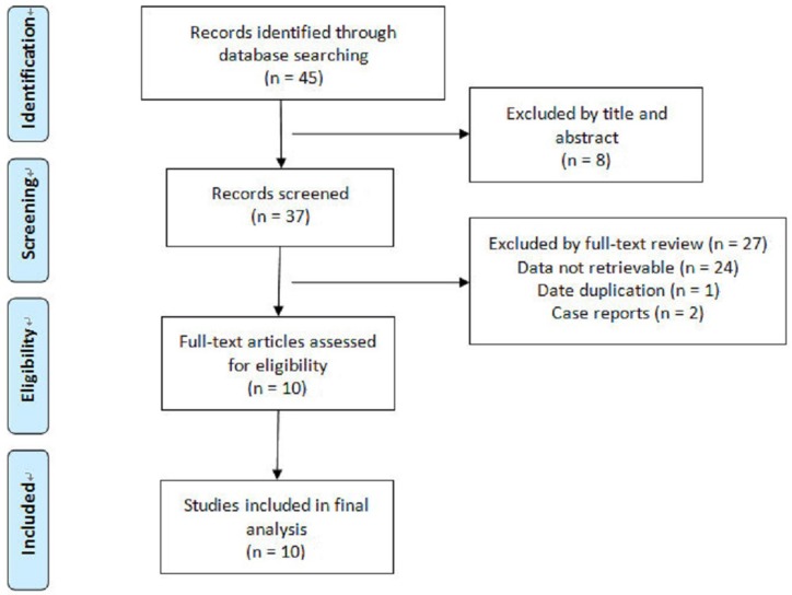
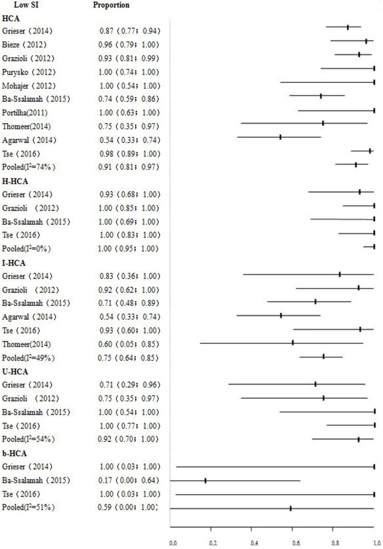
The purpose of this study was to systematically review the diagnostic performance of gadoxetic acid-enhanced magnetic resonance imaging (Gd-EOB-DTPA-MRI) for differentiation of hepatocellular adenoma (HCA) and focal nodular hyperplasia (FNH), as well as HCA classification by using the low signal intensity (SI) in the hepatobiliary phase (HBP). A systematic process was used to review all published data in MEDLINE database about Gd-EOB-DTPA-MRI applied to differentiation of HCA and FNH, and classification of HCA by using low SI in the HBP. The pooled sensitivity and specificity were calculated to assess the diagnostic value of low SI in the HBP. A review of 45 articles identified 10 eligible studies with a total of 288 HCA lesions. The pooled proportion of low SI in the HBP of HCA were 91% (95% CI: 0.81-0.97). In specific, the subtypes of HCA were 75% (95% CI: 0.64-0.85) for I-HCA, 100% (95% CI: 0.95-1.00) for H-HCA, 92% (95% CI: 0.70-1.00) for U-HCA, and 59% (95% CI: 0.00-1.00) for b-HCA, respectively. The pooled specificity and sensitivity of low SI in the HBP for distinguishing FNH from HCA were 95% (95% CI: 0.92-0.98) and 92% (95% CI: 0.87-0.96), respectively. Low SI in the HBP of Gd-EOB-DTPA-MRI is associated with higher accuracy for distinguishing HCA from FNH. However, the diagnostic accuracy may be overvalued, especially for the diagnosis of subtypes of b-HCA and I-HCA. Therefore, the risk factors and conventional imaging findings should be take into account simultaneously.

摘要

本研究旨在系统回顾钆塞酸增强磁共振成像(Gd-EOB-DTPA-MRI)在鉴别肝细胞腺瘤(HCA)和局灶性结节增生(FNH)以及利用肝胆期(HBP)低信号强度(SI)对 HCA 进行分类方面的诊断性能。采用系统的方法检索 MEDLINE 数据库中关于 Gd-EOB-DTPA-MRI 应用于 HCA 和 FNH 鉴别以及利用 HBP 低 SI 对 HCA 进行分类的所有已发表数据。计算汇总的敏感性和特异性,以评估 HBP 低 SI 的诊断价值。对 45 篇文章进行回顾,确定了 10 项符合条件的研究,共 288 个 HCA 病变。HBP 中 HCA 低 SI 的汇总比例为 91%(95%CI:0.81-0.97)。具体而言,HCA 的亚型分别为 I-HCA 75%(95%CI:0.64-0.85)、H-HCA 100%(95%CI:0.95-1.00)、U-HCA 92%(95%CI:0.70-1.00)和 b-HCA 59%(95%CI:0.00-1.00)。HBP 中低 SI 鉴别 FNH 和 HCA 的特异性和敏感性分别为 95%(95%CI:0.92-0.98)和 92%(95%CI:0.87-0.96)。Gd-EOB-DTPA-MRI 的 HBP 中低 SI 与鉴别 HCA 与 FNH 的准确性更高。然而,诊断准确性可能被高估,尤其是对于 b-HCA 和 I-HCA 亚型的诊断。因此,应同时考虑危险因素和常规成像表现。

https://cdn.ncbi.nlm.nih.gov/pmc/blobs/2f14/5562118/1ea8e4b0cca3/ijmsv14p0668g003.jpg
https://cdn.ncbi.nlm.nih.gov/pmc/blobs/2f14/5562118/49f30a0e1f38/ijmsv14p0668g001.jpg
https://cdn.ncbi.nlm.nih.gov/pmc/blobs/2f14/5562118/cd0f4e7083d7/ijmsv14p0668g002.jpg
https://cdn.ncbi.nlm.nih.gov/pmc/blobs/2f14/5562118/1ea8e4b0cca3/ijmsv14p0668g003.jpg
https://cdn.ncbi.nlm.nih.gov/pmc/blobs/2f14/5562118/49f30a0e1f38/ijmsv14p0668g001.jpg
https://cdn.ncbi.nlm.nih.gov/pmc/blobs/2f14/5562118/cd0f4e7083d7/ijmsv14p0668g002.jpg
https://cdn.ncbi.nlm.nih.gov/pmc/blobs/2f14/5562118/1ea8e4b0cca3/ijmsv14p0668g003.jpg

相似文献

1
Diagnostic Value of Gadoxetic Acid-Enhanced MR Imaging to Distinguish HCA and Its Subtype from FNH: A Systematic Review.钆塞酸增强磁共振成像对鉴别 HCA 及其亚型与 FNH 的诊断价值:系统评价。
Int J Med Sci. 2017 Jun 23;14(7):668-674. doi: 10.7150/ijms.17865. eCollection 2017.
2
Hepatic Adenoma Subtypes on Hepatobiliary Phase of Gadoxetic Acid-Enhanced MRI: Systematic Review and Meta-Analysis.钆塞酸二钠增强MRI肝胆期的肝腺瘤亚型:系统评价与Meta分析
AJR Am J Roentgenol. 2023 Jan;220(1):28-38. doi: 10.2214/AJR.22.27989. Epub 2022 Aug 3.
3
The diagnostic value of Gd-EOB-DTPA-MRI for the diagnosis of focal nodular hyperplasia: a systematic review and meta-analysis.钆塞酸二钠增强磁共振成像对肝脏局灶性结节增生的诊断价值:一项系统评价与Meta分析
Eur Radiol. 2015 Apr;25(4):950-60. doi: 10.1007/s00330-014-3499-9. Epub 2014 Dec 24.
4
Intrapatient Comparison of the Hepatobiliary Phase of Gd-BOPTA and Gd-EOB-DTPA in the Differentiation of Hepatocellular Adenoma From Focal Nodular Hyperplasia.患者内比较 Gd-BOPTA 和 Gd-EOB-DTPA 的肝胆期在肝细胞腺瘤与局灶性结节增生鉴别诊断中的作用。
J Magn Reson Imaging. 2019 Mar;49(3):700-710. doi: 10.1002/jmri.26227. Epub 2018 Sep 25.
5
OATPB1/B3 and MRP3 expression in hepatocellular adenoma predicts Gd-EOB-DTPA uptake and correlates with risk of malignancy.OATPB1/B3 和 MRP3 在肝细胞腺瘤中的表达可预测 Gd-EOB-DTPA 摄取,与恶性风险相关。
Liver Int. 2019 Jan;39(1):158-167. doi: 10.1111/liv.13964. Epub 2018 Oct 8.
6
Gadoxetic acid-enhanced MRI in differentiating focal nodular hyperplasia from hepatocellular adenoma in children.钆塞酸二钠增强磁共振成像在鉴别儿童局灶性结节性增生与肝细胞腺瘤中的应用
Br J Radiol. 2025 Feb 1;98(1166):262-270. doi: 10.1093/bjr/tqae222.
7
HBP-enhancing hepatocellular adenomas and how to discriminate them from FNH in Gd-EOB MRI.增强型肝细胞腺瘤及其在钆塞酸增强 MRI 中的与 FNH 鉴别
BMC Med Imaging. 2021 Feb 15;21(1):28. doi: 10.1186/s12880-021-00552-0.
8
Diagnostic value of morphological enhancement patterns in the hepatobiliary phase of gadoxetic acid-enhanced MRI to distinguish focal nodular hyperplasia from hepatocellular adenoma.钆塞酸增强 MRI 肝胆期形态增强模式对鉴别局灶性结节增生与肝细胞腺瘤的诊断价值。
Radiol Med. 2021 Nov;126(11):1379-1387. doi: 10.1007/s11547-021-01403-2. Epub 2021 Jul 21.
9
[Gd-EOB-DTPA-enhanced magnetic resonance imaging: differentiation between focal nodular hyperplasia and hepatocellular adenoma].[钆塞酸二钠增强磁共振成像:局灶性结节性增生与肝细胞腺瘤的鉴别诊断]
Acta Med Port. 2011 Dec;24 Suppl 2:531-8. Epub 2011 Dec 31.
10
Lesions hyper- to isointense to surrounding liver in the hepatobiliary phase of gadoxetic acid-enhanced MRI.在钆塞酸增强 MRI 的肝胆期,病灶呈高于或等于周围肝脏的信号强度。
Eur Radiol. 2024 Dec;34(12):7661-7672. doi: 10.1007/s00330-024-10829-x. Epub 2024 Jun 20.

引用本文的文献

1
The value of Gd-EOB-DTPA-enhanced MRI in the differential diagnosis of HCC with hyperintensity on HBP and FNH.钆塞酸二钠增强磁共振成像在肝细胞癌与肝胆期高信号的局灶性结节性增生鉴别诊断中的价值
Medicine (Baltimore). 2025 Jun 27;104(26):e43106. doi: 10.1097/MD.0000000000043106.
2
Practical approach to diagnose and manage benign liver masses.诊断和处理良性肝肿块的实用方法。
Hepatol Commun. 2024 Oct 30;8(11). doi: 10.1097/HC9.0000000000000560. eCollection 2024 Nov 1.
3
Hepatocellular adenoma update: diagnosis, molecular classification, and clinical course.

本文引用的文献

1
Qualitative and Quantitative Gadoxetic Acid-enhanced MR Imaging Helps Subtype Hepatocellular Adenomas.定性和定量钆塞酸增强磁共振成像有助于肝腺瘤亚型分类。
Radiology. 2016 Apr;279(1):118-27. doi: 10.1148/radiol.2015142449. Epub 2015 Oct 27.
2
Morphologic and Molecular Features of Hepatocellular Adenoma with Gadoxetic Acid-enhanced MR Imaging.钆塞酸增强磁共振成像在肝细胞腺瘤中的形态学和分子特征。
Radiology. 2015 Oct;277(1):104-13. doi: 10.1148/radiol.2015142366. Epub 2015 May 18.
3
Hepatocellular adenoma management: call for shared guidelines and multidisciplinary approach.
肝细胞腺瘤更新:诊断、分子分类和临床过程。
Br J Radiol. 2024 Nov 1;97(1163):1740-1754. doi: 10.1093/bjr/tqae180.
4
Lesions hyper- to isointense to surrounding liver in the hepatobiliary phase of gadoxetic acid-enhanced MRI.在钆塞酸增强 MRI 的肝胆期,病灶呈高于或等于周围肝脏的信号强度。
Eur Radiol. 2024 Dec;34(12):7661-7672. doi: 10.1007/s00330-024-10829-x. Epub 2024 Jun 20.
5
Diagnostic Values of the Liver Imaging Reporting and Data System in the Detection and Characterization of Hepatocellular Carcinoma: A Systematic Review and Meta-Analysis.肝脏影像报告和数据系统在肝细胞癌检测与特征描述中的诊断价值:一项系统评价和Meta分析
Cureus. 2023 Mar 13;15(3):e36082. doi: 10.7759/cureus.36082. eCollection 2023 Mar.
6
β-Catenin-activated inflammatory hepatocellular adenoma with pigmentation and atypical features: a case report.伴有色素沉着和非典型特征的β-连环蛋白激活型炎性肝细胞腺瘤:一例报告
Clin J Gastroenterol. 2023 Apr;16(2):237-243. doi: 10.1007/s12328-023-01757-2. Epub 2023 Jan 14.
7
Hepatobiliary phases in magnetic resonance imaging using liver-specific contrast for focal lesions in clinical practice.临床实践中使用肝脏特异性对比剂对局灶性病变进行磁共振成像的肝胆期。
World J Hepatol. 2022 Jul 27;14(7):1459-1469. doi: 10.4254/wjh.v14.i7.1459.
8
Hepatic Adenoma Subtypes on Hepatobiliary Phase of Gadoxetic Acid-Enhanced MRI: Systematic Review and Meta-Analysis.钆塞酸二钠增强MRI肝胆期的肝腺瘤亚型:系统评价与Meta分析
AJR Am J Roentgenol. 2023 Jan;220(1):28-38. doi: 10.2214/AJR.22.27989. Epub 2022 Aug 3.
9
A Scoping Review of the Classification, Diagnosis, and Management of Hepatic Adenomas.肝腺瘤分类、诊断及管理的范围综述
J Gastrointest Surg. 2022 Apr;26(4):965-978. doi: 10.1007/s11605-022-05246-8. Epub 2022 Jan 26.
10
Application of Immunohistochemistry in the Pathological Diagnosis of Liver Tumors.免疫组织化学在肝脏肿瘤病理诊断中的应用。
Int J Mol Sci. 2021 May 28;22(11):5780. doi: 10.3390/ijms22115780.
肝细胞腺瘤的管理:呼吁制定共享指南和采取多学科方法。
Clin Res Hepatol Gastroenterol. 2015 Apr;39(2):180-7. doi: 10.1016/j.clinre.2014.10.003. Epub 2014 Nov 26.
4
Inflammatory hepatocellular adenomas can mimic focal nodular hyperplasia on gadoxetic acid-enhanced MRI.炎症性肝细胞腺瘤在钆塞酸增强磁共振成像(MRI)上可模仿局灶性结节性增生。
AJR Am J Roentgenol. 2014 Oct;203(4):W408-14. doi: 10.2214/AJR.13.12251. Epub 2014 Jul 23.
5
Gadoxetic acid enhanced MRI for differentiation of FNH and HCA: a single centre experience.钆塞酸增强 MRI 鉴别 FNH 和 HCA:单中心经验。
Eur Radiol. 2014 Jun;24(6):1339-48. doi: 10.1007/s00330-014-3144-7.
6
Genotype-phenotype correlations in hepatocellular adenoma: an update of MRI findings.肝细胞腺瘤的基因型-表型相关性:MRI 表现的最新研究进展。
Diagn Interv Radiol. 2014 May-Jun;20(3):193-9. doi: 10.5152/dir.2013.13315.
7
[Gd-EOB-DTPA-enhanced magnetic resonance imaging: differentiation between focal nodular hyperplasia and hepatocellular adenoma].[钆塞酸二钠增强磁共振成像:局灶性结节性增生与肝细胞腺瘤的鉴别诊断]
Acta Med Port. 2011 Dec;24 Suppl 2:531-8. Epub 2011 Dec 31.
8
Diagnostic accuracy of MRI in differentiating hepatocellular adenoma from focal nodular hyperplasia: prospective study of the additional value of gadoxetate disodium.MRI 对肝细胞腺瘤与局灶性结节增生的诊断准确性:钆塞酸二钠的附加价值的前瞻性研究。
AJR Am J Roentgenol. 2012 Jul;199(1):26-34. doi: 10.2214/AJR.11.7750.
9
Characterization of hepatic adenoma and focal nodular hyperplasia with gadoxetic acid.钆塞酸增强磁共振成像对肝腺瘤和局灶性结节增生的特征分析。
J Magn Reson Imaging. 2012 Sep;36(3):686-96. doi: 10.1002/jmri.23701. Epub 2012 Jun 4.
10
Systematic review of haemorrhage and rupture of hepatocellular adenomas.系统综述肝细胞腺瘤的出血和破裂。
Br J Surg. 2012 Jul;99(7):911-6. doi: 10.1002/bjs.8762. Epub 2012 May 22.