Massignan Ângela, Wagner Felipe Victora, Toniolo de Carvalho Pedro, da Silveira Cima Alexandre
Department of Radiology, Hospital Moinhos de Vento, Ramiro Barcelos, 910, Moinhos de Vento, Porto Alegre, Rio Grande do Sul 90035-001, Brazil.
Radiol Case Rep. 2017 Jun 10;12(3):577-584. doi: 10.1016/j.radcr.2017.04.015. eCollection 2017 Sep.
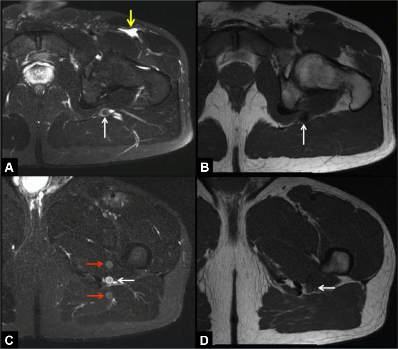
The persistent sciatic artery is a rare anatomical variant, representing the persistence of the sciatic artery in adult life that is responsible for the major blood supply to the lower limb in early embryologic development. Such persistence may be bilateral and can remain asymptomatic for many years. However, aneurysmal degeneration has been described as a complication of the persistent sciatic artery, which may cause critical limb ischemia resulting from thrombosis or embolization of aneurysm thrombus. Digital subtraction angiography, Doppler ultrasound, computed tomography angiography and magnetic resonance angiography are the most frequently used diagnostic tools to detect, classify and determine the presence of complications of a PSA. Early detection of this vascular abnormality on imaging studies can avoid life-threatening complications. We describe 4 patients with PSA that were diagnosed as an incidental finding in magnetic resonance imaging of the hip and demonstrate its characteristic imaging appearance.
坐骨动脉持续存在是一种罕见的解剖变异,指在成年期坐骨动脉持续存在,而在胚胎发育早期它是下肢主要的血液供应来源。这种持续存在可能是双侧的,并且多年来可能一直无症状。然而,动脉瘤样退变已被描述为坐骨动脉持续存在的一种并发症,这可能导致因动脉瘤血栓形成血栓或栓塞而引起严重肢体缺血。数字减影血管造影、多普勒超声、计算机断层血管造影和磁共振血管造影是检测、分类和确定坐骨动脉持续存在并发症的最常用诊断工具。在影像学检查中早期发现这种血管异常可避免危及生命的并发症。我们描述了4例在髋关节磁共振成像中偶然发现的坐骨动脉持续存在病例,并展示了其特征性的影像学表现。