Department of Internal Medicine, Gastroenterology Unit, University of Modena and Reggio Emilia, Modena, Italy.
WomenInHepatology Network, Modena, Italy.
Cell Death Dis. 2017 Aug 24;8(8):e3017. doi: 10.1038/cddis.2017.395.
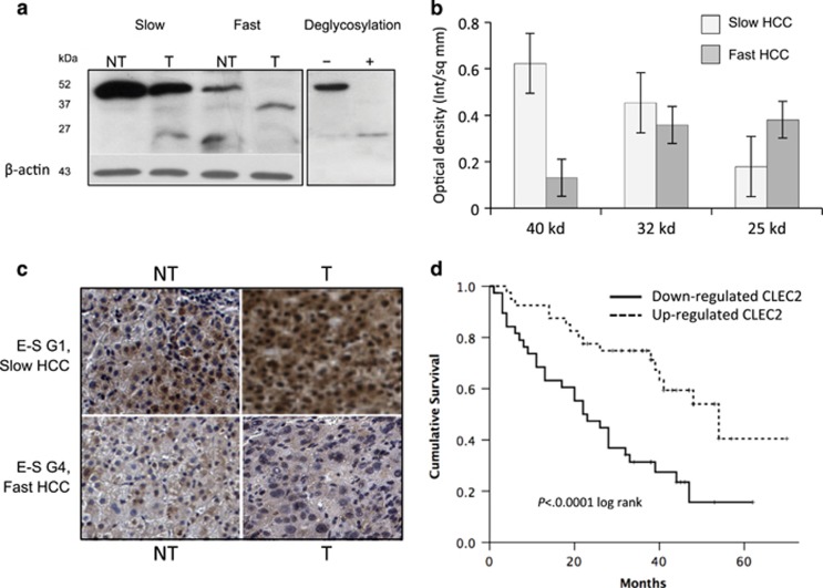
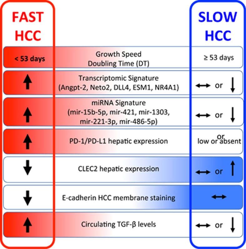
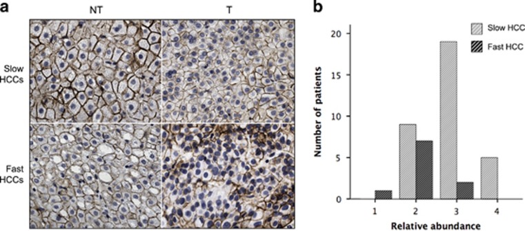
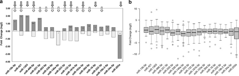
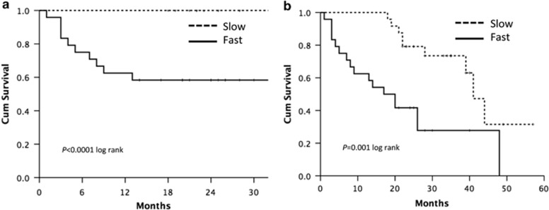
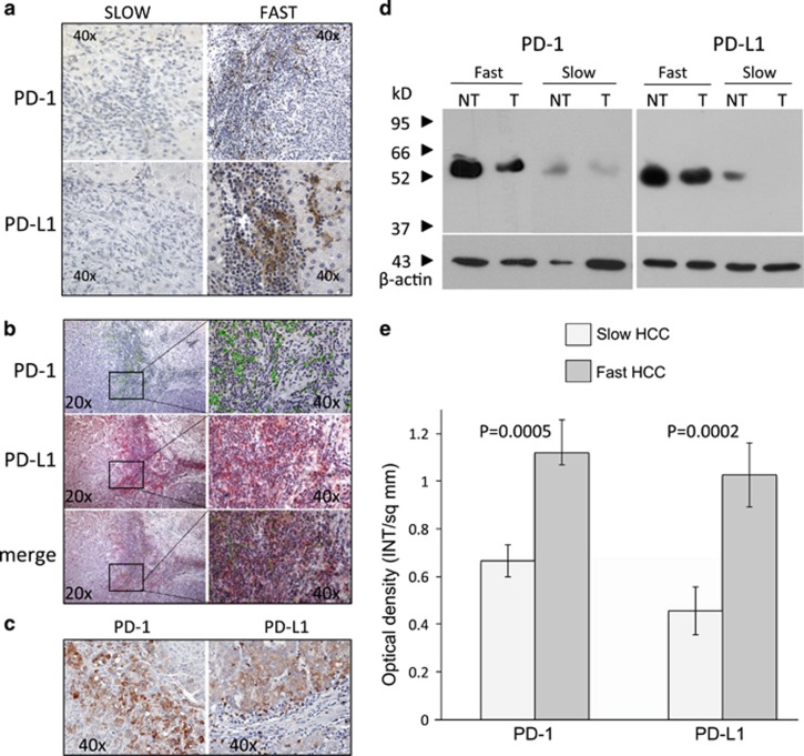
In HCC, tumor microenvironment, heavily influenced by the underlying chronic liver disease, etiology and stage of the tissue damage, affects tumor progression and determines the high heterogeneity of the tumor. Aim of this study was to identify the circulating and tissue components of the microenvironment immune-mediated response affecting the aggressiveness and the ensuing clinical outcome. We analyzed the baseline paired HCC and the surrounding tissue biopsies from a prospective cohort of 132 patients at the first diagnosis of HCC for immunolocalization of PD-1/PD-L1, FoxP3, E-cadherin, CLEC2 and for a panel of 82 microRNA associated with regulation of angiogenesis, cell proliferation, cell signaling, immune control and autophagy. Original microarray data were also explored. Serum samples were analyzed for a panel of 19 cytokines. Data were associated with biochemical data, histopathology and survival. Patients with a more aggressive disease and shorter survival, who we named fast-growing accordingly to the tumor doubling time, at presentation had significantly higher AFP levels, TGF-β1 and Cyphra 21-1 levels. Transcriptomic analysis evidenced a significant downregulation of CLEC2 and upregulation of several metalloproteinases. A marked local upregulation of both PD-1 and PD-L1, a concomitant FoxP3-positive lymphocytic infiltrate, a loss of E-cadherin, gain of epithelial-mesenchymal transition (EMT) phenotype and extreme poor differentiation at histology were also present. Upregulated microRNA in fast-growing HCCs are associated with TGF-β signaling, angiogenesis and inflammation. Our data show that fast HCCs are characterized not only by redundant neo-angiogenesis but also by unique features of distinctively immunosuppressed microenvironment, prominent EMT, and clear-cut activation of TGFβ1 signaling in a general background of long-standing and permanent inflammatory state.
在 HCC 中,肿瘤微环境受潜在慢性肝病、病因和组织损伤阶段的严重影响,会影响肿瘤的进展并决定肿瘤的高度异质性。本研究旨在确定影响侵袭性和后续临床结果的免疫介导的微环境循环和组织成分。我们分析了 132 例 HCC 患者的前瞻性队列中基线配对的 HCC 和周围组织活检,对 PD-1/PD-L1、FoxP3、E-钙黏蛋白、CLEC2 进行免疫定位,并对与血管生成、细胞增殖、细胞信号转导、免疫控制和自噬调节相关的 82 个 microRNA 进行了分析。还探索了原始微阵列数据。分析了血清样本中与一组 19 种细胞因子相关的信息。数据与生化数据、组织病理学和生存情况相关联。表现出侵袭性更强和生存时间更短的患者,我们根据肿瘤倍增时间将其命名为快速生长型,他们的 AFP 水平、TGF-β1 和 Cyphra 21-1 水平显著更高。转录组分析表明 CLEC2 的表达显著下调,而几种金属蛋白酶的表达显著上调。局部 PD-1 和 PD-L1 的表达明显上调,伴有 FoxP3 阳性淋巴细胞浸润,E-钙黏蛋白丢失,上皮-间充质转化(EMT)表型获得,组织学上分化极差。快速生长型 HCC 中上调的 microRNA 与 TGF-β 信号、血管生成和炎症有关。我们的数据表明,快速生长的 HCC 不仅具有冗余的新生血管生成,而且具有独特的免疫抑制微环境特征,突出的 EMT,以及在长期和永久性炎症状态的普遍背景下明显激活 TGFβ1 信号。