Christoph F Dietrich, Medizinische Klinik 2, Caritas-Krankenhaus Bad Mergentheim, 97980 Bad Mergentheim, Germany.
World J Gastroenterol. 2017 Aug 14;23(30):5567-5578. doi: 10.3748/wjg.v23.i30.5567.
To describe the imaging features of serous neoplasms of the pancreas using ultrasound, endoscopic ultrasound, computed tomography and magnetic resonance imaging.
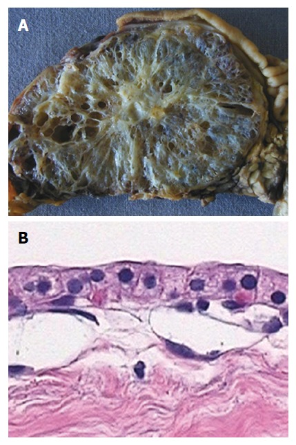
This multicenter international collaboration enhances a literature review to date, reporting features of 287 histologically confirmed cases of serous pancreatic cystic neoplasms (SPNs).
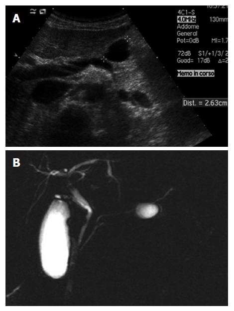
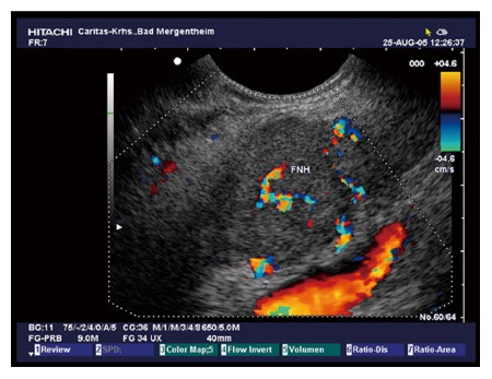
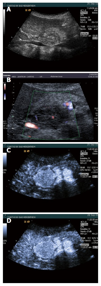
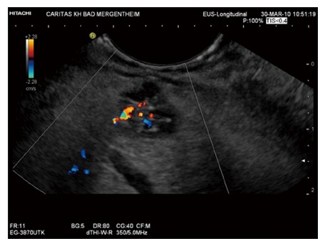
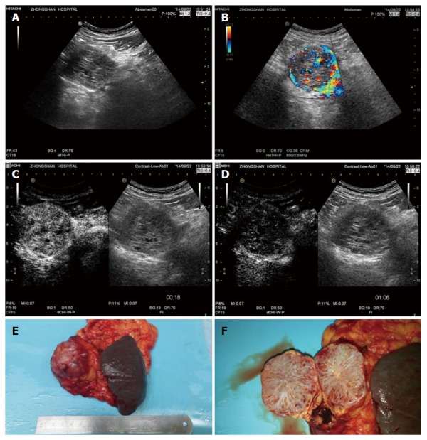
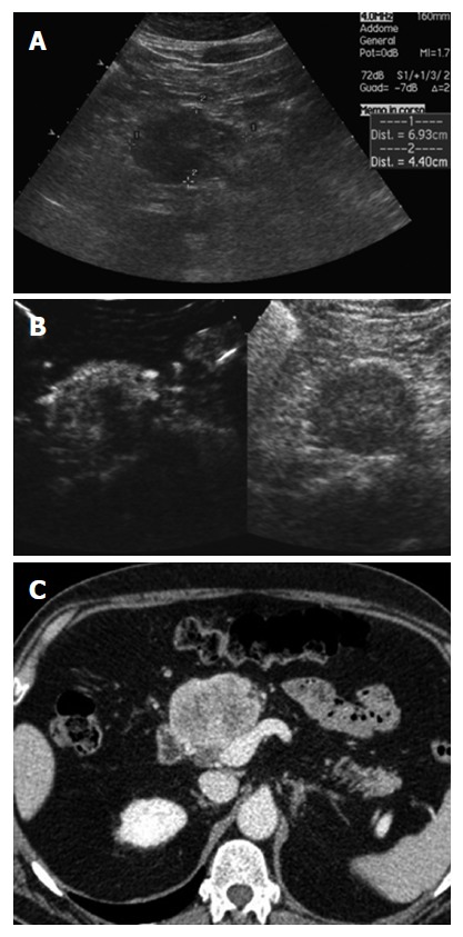
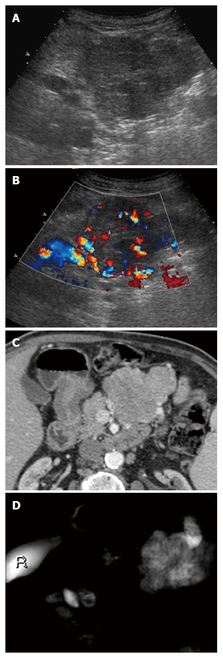
Female predominance is seen with most SPNs presenting asymptomatically in the 5 through 7 decade. Mean lesion size was 38.7 mm, 98% were single, 44.2% cystic, 46% mixed cystic and solid, and 94% hypoechoic on B-mode ultrasound. Vascular patterns and contrast-enhancement profiles are described as hypervascular and hyperenhancing.
The described ultrasound features can aid differentiation of SPN from other neoplastic lesions under most circumstances.
使用超声、内镜超声、计算机断层扫描和磁共振成像描述胰腺浆液性肿瘤的影像学特征。
本多中心国际合作在迄今为止的文献综述的基础上进行了扩展,报告了 287 例经组织学证实的胰腺浆液性囊性肿瘤(SPN)的特征。
大多数 SPN 以女性为主,无症状表现,发病年龄在 5 至 7 十年代。平均病变大小为 38.7 毫米,98%为单发,44.2%为囊性,46%为囊实性混合,94%在 B 模式超声下呈低回声。描述了血管模式和对比增强特征为富血管性和高增强性。
在大多数情况下,描述的超声特征可以帮助区分 SPN 与其他肿瘤性病变。