• 文献检索
  • 文档翻译
  • 深度研究
  • 学术资讯
  • Suppr Zotero 插件Zotero 插件
  • 邀请有礼
  • 套餐&价格
  • 历史记录
应用&插件
Suppr Zotero 插件Zotero 插件浏览器插件Mac 客户端Windows 客户端微信小程序
定价
高级版会员购买积分包购买API积分包
服务
文献检索文档翻译深度研究API 文档MCP 服务
关于我们
关于 Suppr公司介绍联系我们用户协议隐私条款
关注我们

Suppr 超能文献

核心技术专利:CN118964589B侵权必究
粤ICP备2023148730 号-1Suppr @ 2026

文献检索

告别复杂PubMed语法,用中文像聊天一样搜索,搜遍4000万医学文献。AI智能推荐,让科研检索更轻松。

立即免费搜索

文件翻译

保留排版,准确专业,支持PDF/Word/PPT等文件格式,支持 12+语言互译。

免费翻译文档

深度研究

AI帮你快速写综述,25分钟生成高质量综述,智能提取关键信息,辅助科研写作。

立即免费体验

应变直方图超声内镜弹性成像和对比增强超声内镜在胰腺局灶性肿块鉴别诊断中的临床应用:一项前瞻性多中心研究。

Clinical impact of strain histogram EUS elastography and contrast-enhanced EUS for the differential diagnosis of focal pancreatic masses: A prospective multicentric study.

作者信息

Costache Mădălin Ionuţ, Cazacu Irina M, Dietrich Christoph F, Petrone Maria Chiara, Arcidiacono Paolo Giorgio, Giovannini Marc, Bories Erwan, Garcia Julio Iglesias, Siyu Sun, Santo Erwin, Popescu Carmen Florina, Constantin Alina, Bhutani Manoop S, Saftoiu Adrian

机构信息

Research Center of Gastroenterology and Hepatology, University of Medicine and Pharmacy, Craiova, Romania.

Research Center of Gastroenterology and Hepatology, University of Medicine and Pharmacy, Craiova, Romania; Department of Gastroenterology, Hepatology and Nutrition, MD Anderson Cancer Center, Houston, Texas, USA.

出版信息

Endosc Ultrasound. 2020 Mar-Apr;9(2):116-121. doi: 10.4103/eus.eus_69_19.

DOI:10.4103/eus.eus_69_19
PMID:32295969
原文链接:https://pmc.ncbi.nlm.nih.gov/articles/PMC7279079/
Abstract

BACKGROUND

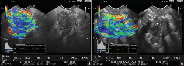
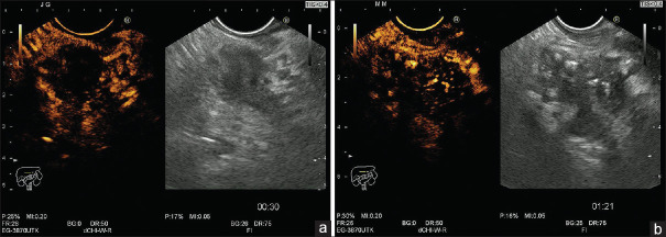
Recent advances in EUS techniques (real-time EUS elastography and contrast-enhanced EUS) have allowed a better characterization of focal pancreatic masses. Mean strain histograms (SHs) are considered a good parameter for the semi-quantitative evaluation of focal pancreatic masses, alongside complementary contrast-enhanced EUS parameters which can be quantified during both the early arterial and late venous phase.

MATERIALS AND METHODS

The study design was prospective, blinded, and multicentric, assessing real-time EUS elastography and contrast-enhanced EUS results for the characterization of focal pancreatic masses using parametric measurements, in comparison with pathology which is the gold standard. SHs were performed based on the embedded software of the ultrasound system, with the values being reversed as opposed to our initially published data on hue histograms. Consequently, a cutoff of 80 was derived from previous multicentric trials. Contrast-enhanced EUS also allowed the focal masses to be classified as hyper-, iso-, or hypoenhanced in comparison with the normal pancreatic parenchyma. EUS-FNA was then performed for all patients, with a positive cytological diagnosis taken as a final proof of malignancy for the pancreatic masses. The diagnoses obtained by EUS-FNA were verified further either by surgery or during a clinical follow-up of at least 6 months.

RESULTS

A total number of 97 consecutive patients with focal pancreatic masses were included in the study. Based on previously defined cutoffs of 80, the values of sensitivity, specificity, positive predictive value, negative predictive value, and accuracy of the mean SHs for the diagnosis of pancreatic cancer were 100%, 29.63%, 78.65%, 100%, and 80.41%, respectively. Corresponding values for contrast-enhanced EUS (taking into consideration hypoenhencement as a predictive factor of malignancy) were 98.57%, 77.78%, 92%, 95.45%, and 92.78%, respectively. Combining contrast enhancement-EUS (hypoenhencement) and semi-quantitative EUS elastography (SH cutoffs <80), the resulting values corresponding for sensitivity, specificity, and accuracy were 98.57%, 81.48%, and 93.81%, respectively.

CONCLUSION

The current study using objective parametric tools for both EUS elastography and contrast-enhanced EUS confirmed the results of previous studies and meta-analyses that indicated a complementary role for the differential diagnosis of focal pancreatic masses. Moreover, the best values for the receiver operating curves were obtained using a sequential clinical algorithm based on the initial use of elastography, followed by contrast enhancement.

摘要

背景

超声内镜(EUS)技术(实时EUS弹性成像和对比增强EUS)的最新进展使胰腺局灶性肿块的特征得以更好地描述。平均应变直方图(SHs)被认为是胰腺局灶性肿块半定量评估的一个良好参数,同时还有可在动脉早期和静脉晚期进行量化的对比增强EUS补充参数。

材料与方法

本研究设计为前瞻性、盲法、多中心研究,通过参数测量评估实时EUS弹性成像和对比增强EUS结果以描述胰腺局灶性肿块特征,并与作为金标准的病理结果进行比较。SHs根据超声系统的嵌入式软件进行测量,与我们最初发表的关于色调直方图的数据相反,其值进行了反转。因此,根据之前的多中心试验得出截断值为80。对比增强EUS还可将局灶性肿块与正常胰腺实质相比分为高增强、等增强或低增强。然后对所有患者进行EUS-FNA,将阳性细胞学诊断作为胰腺肿块恶性肿瘤的最终确诊依据。通过手术或至少6个月的临床随访进一步验证EUS-FNA获得的诊断结果。

结果

本研究共纳入97例连续的胰腺局灶性肿块患者。根据之前定义的截断值80,平均SHs诊断胰腺癌的敏感性、特异性、阳性预测值、阴性预测值和准确性分别为100%、29.63%、78.65%、100%和80.41%。对比增强EUS(将低增强作为恶性肿瘤的预测因素)的相应值分别为98.57%、77.78%、92%、95.45%和92.78%。将对比增强EUS(低增强)和半定量EUS弹性成像(SH截断值<80)相结合,敏感性、特异性和准确性的相应值分别为98.57%、81.48%和93.81%。

结论

本研究使用针对EUS弹性成像和对比增强EUS的客观参数工具,证实了之前研究和荟萃分析的结果,即两者在胰腺局灶性肿块的鉴别诊断中具有互补作用。此外,基于最初使用弹性成像,随后进行对比增强的序贯临床算法获得了最佳的受试者工作曲线值。

https://cdn.ncbi.nlm.nih.gov/pmc/blobs/3f0b/7279079/56ba9f8413e8/EUS-9-116-g002.jpg
https://cdn.ncbi.nlm.nih.gov/pmc/blobs/3f0b/7279079/af2c2e01a8f0/EUS-9-116-g001.jpg
https://cdn.ncbi.nlm.nih.gov/pmc/blobs/3f0b/7279079/56ba9f8413e8/EUS-9-116-g002.jpg
https://cdn.ncbi.nlm.nih.gov/pmc/blobs/3f0b/7279079/af2c2e01a8f0/EUS-9-116-g001.jpg
https://cdn.ncbi.nlm.nih.gov/pmc/blobs/3f0b/7279079/56ba9f8413e8/EUS-9-116-g002.jpg

相似文献

1
Clinical impact of strain histogram EUS elastography and contrast-enhanced EUS for the differential diagnosis of focal pancreatic masses: A prospective multicentric study.应变直方图超声内镜弹性成像和对比增强超声内镜在胰腺局灶性肿块鉴别诊断中的临床应用:一项前瞻性多中心研究。
Endosc Ultrasound. 2020 Mar-Apr;9(2):116-121. doi: 10.4103/eus.eus_69_19.
2
Neural network analysis of dynamic sequences of EUS elastography used for the differential diagnosis of chronic pancreatitis and pancreatic cancer.用于慢性胰腺炎和胰腺癌鉴别诊断的超声内镜弹性成像动态序列的神经网络分析
Gastrointest Endosc. 2008 Dec;68(6):1086-94. doi: 10.1016/j.gie.2008.04.031. Epub 2008 Jul 24.
3
Clinical impact of EUS elastography followed by contrast-enhanced EUS in patients with focal pancreatic masses and negative EUS-guided FNA.超声内镜弹性成像联合超声内镜造影在胰腺局灶性肿块且超声内镜引导下细针穿刺活检阴性患者中的临床影响
Med Ultrason. 2016 Mar;18(1):18-24. doi: 10.11152/mu.2013.2066.181.ich.
4
Endoscopic ultrasound elastography strain histograms in the evaluation of patients with pancreatic masses.内镜超声弹性成像应变直方图在胰腺肿块患者评估中的应用
World J Gastroenterol. 2015 Apr 7;21(13):4014-9. doi: 10.3748/wjg.v21.i13.4014.
5
Combined contrast-enhanced power Doppler and real-time sonoelastography performed during EUS, used in the differential diagnosis of focal pancreatic masses (with videos).超声内镜检查时联合使用增强能量多普勒和实时超声弹性成像进行诊断,用于鉴别诊断局灶性胰腺肿块(附有视频)。
Gastrointest Endosc. 2010 Oct;72(4):739-47. doi: 10.1016/j.gie.2010.02.056. Epub 2010 Aug 2.
6
Quantitative low mechanical index contrast-enhanced endoscopic ultrasound for the differential diagnosis of chronic pseudotumoral pancreatitis and pancreatic cancer.定量低机械指数对比增强内镜超声在慢性假性肿瘤性胰腺炎和胰腺癌鉴别诊断中的应用。
BMC Gastroenterol. 2013 Jan 3;13:2. doi: 10.1186/1471-230X-13-2.
7
Efficacy of an artificial neural network-based approach to endoscopic ultrasound elastography in diagnosis of focal pancreatic masses.基于人工神经网络的内镜超声弹性成像在诊断局灶性胰腺肿块中的效能。
Clin Gastroenterol Hepatol. 2012 Jan;10(1):84-90.e1. doi: 10.1016/j.cgh.2011.09.014. Epub 2011 Oct 1.
8
Accuracy of endoscopic ultrasound elastography used for differential diagnosis of focal pancreatic masses: a multicenter study.内镜超声弹性成像在局灶性胰腺肿块鉴别诊断中的准确性:一项多中心研究。
Endoscopy. 2011 Jul;43(7):596-603. doi: 10.1055/s-0030-1256314. Epub 2011 Mar 24.
9
Diagnostic accuracy of quantitative EUS elastography for discriminating malignant from benign solid pancreatic masses: a prospective, single-center study.定量超声弹性成像诊断胰腺实性肿块良恶性的准确性:一项前瞻性单中心研究。
Gastrointest Endosc. 2012 Nov;76(5):953-61. doi: 10.1016/j.gie.2012.05.034. Epub 2012 Jul 31.
10
Quantitative endoscopic ultrasound elastography: an accurate method for the differentiation of solid pancreatic masses.定量内镜超声弹性成像:一种准确区分胰腺实性肿块的方法。
Gastroenterology. 2010 Oct;139(4):1172-80. doi: 10.1053/j.gastro.2010.06.059. Epub 2010 Jun 27.

引用本文的文献

1
Comparison of standard auxiliary (contrast or elastography) endoscopic ultrasound-guided fine needle aspiration/biopsy in solid pancreatic lesions: A meta-analysis.标准辅助(对比或弹性成像)内镜超声引导下细针穿刺抽吸/活检在实性胰腺病变中的比较:一项荟萃分析。
World J Methodol. 2025 Sep 20;15(3):97415. doi: 10.5662/wjm.v15.i3.97415.
2
EUS-based intratumoral and peritumoral machine learning radiomics analysis for distinguishing pancreatic neuroendocrine tumors from pancreatic cancer.基于超声内镜的肿瘤内及肿瘤周围机器学习影像组学分析,用于鉴别胰腺神经内分泌肿瘤与胰腺癌。
Front Oncol. 2025 Mar 4;15:1442209. doi: 10.3389/fonc.2025.1442209. eCollection 2025.
3

本文引用的文献

1
Endoscopic ultrasound elastography in the diagnosis of pancreatic masses: A meta-analysis.内镜超声弹性成像在胰腺肿块诊断中的应用:一项荟萃分析。
Pancreatology. 2018 Oct;18(7):833-840. doi: 10.1016/j.pan.2018.07.008. Epub 2018 Jul 30.
2
A quarter century of EUS-FNA: Progress, milestones, and future directions.超声内镜引导下细针穿刺活检25年:进展、里程碑与未来方向
Endosc Ultrasound. 2018 May-Jun;7(3):141-160. doi: 10.4103/eus.eus_19_18.
3
Endoscopic ultrasound elastography of small solid pancreatic lesions: a multicenter study.小胰腺实体病灶的内镜超声弹性成像:一项多中心研究。
Endoscopic ultrasonography-based intratumoral and peritumoral machine learning ultrasomics model for predicting the pathological grading of pancreatic neuroendocrine tumors.
基于内镜超声的肿瘤内和肿瘤周围机器学习超声组学模型用于预测胰腺神经内分泌肿瘤的病理分级
BMC Med Imaging. 2025 Jan 18;25(1):22. doi: 10.1186/s12880-025-01555-x.
4
Endoscopic Ultrasound-Guided Pancreatic Tissue Sampling: Lesion Assessment, Needles, and Techniques.内镜超声引导下胰腺组织采样:病变评估、穿刺针及技术
Medicina (Kaunas). 2024 Dec 7;60(12):2021. doi: 10.3390/medicina60122021.
5
A novel endoscopic ultrasomics-based machine learning model and nomogram to predict the pathological grading of pancreatic neuroendocrine tumors.一种基于新型内镜超声组学的机器学习模型和列线图,用于预测胰腺神经内分泌肿瘤的病理分级。
Heliyon. 2024 Jul 9;10(14):e34344. doi: 10.1016/j.heliyon.2024.e34344. eCollection 2024 Jul 30.
6
Endoscopic ultrasonography-based intratumoral and peritumoral machine learning radiomics analyses for distinguishing insulinomas from non-functional pancreatic neuroendocrine tumors.基于内镜超声的肿瘤内和肿瘤周围机器学习放射组学分析,用于鉴别胰岛素瘤与无功能性胰腺神经内分泌肿瘤。
Front Endocrinol (Lausanne). 2024 Jun 17;15:1383814. doi: 10.3389/fendo.2024.1383814. eCollection 2024.
7
Construction and validation of an endoscopic ultrasonography-based ultrasomics nomogram for differentiating pancreatic neuroendocrine tumors from pancreatic cancer.基于内镜超声的超声组学列线图用于鉴别胰腺神经内分泌肿瘤与胰腺癌的构建与验证
Front Oncol. 2024 May 23;14:1359364. doi: 10.3389/fonc.2024.1359364. eCollection 2024.
8
Diagnostic Endoscopic Ultrasound in Pancreatology: Focus on Normal Variants and Pancreatic Masses.胰腺病学中的诊断性内镜超声:聚焦于正常变异和胰腺肿物
Visc Med. 2023 Oct;39(5):121-130. doi: 10.1159/000533432. Epub 2023 Sep 5.
9
A review of contrast-enhanced harmonic endoscopic ultrasonography for pancreatic solid tumors.对比增强谐波内镜超声检查在胰腺实性肿瘤中的应用综述
J Med Ultrason (2001). 2023 Aug 16. doi: 10.1007/s10396-023-01346-3.
10
The Latest Advancements in Diagnostic Role of Endosonography of Pancreatic Lesions.胰腺病变内镜超声诊断作用的最新进展
J Clin Med. 2023 Jul 12;12(14):4630. doi: 10.3390/jcm12144630.
Endoscopy. 2018 Nov;50(11):1071-1079. doi: 10.1055/a-0588-4941. Epub 2018 Apr 24.
4
EUS elastography: How to do it?超声内镜弹性成像:如何操作?
Endosc Ultrasound. 2018 Jan-Feb;7(1):20-28. doi: 10.4103/eus.eus_49_17.
5
Technique of qualitative and semiquantitative EUS elastography in pancreatic examination.胰腺检查中定性和半定量超声内镜弹性成像技术
Endosc Ultrasound. 2017 Dec;6(Suppl 3):S111-S114. doi: 10.4103/eus.eus_75_17.
6
Strain Elastography - How To Do It?应变弹性成像——如何操作?
Ultrasound Int Open. 2017 Sep;3(4):E137-E149. doi: 10.1055/s-0043-119412. Epub 2017 Dec 7.
7
Present status and perspectives of endosonography 2017 in gastroenterology.2017年超声内镜在胃肠病学中的现状与展望
Korean J Intern Med. 2018 Jan;33(1):36-63. doi: 10.3904/kjim.2017.212. Epub 2017 Nov 23.
8
Serous pancreatic neoplasia, data and review.浆液性胰腺肿瘤,数据与综述。
World J Gastroenterol. 2017 Aug 14;23(30):5567-5578. doi: 10.3748/wjg.v23.i30.5567.
9
Autoimmune pancreatitis: Imaging features.自身免疫性胰腺炎:影像学特征。
Endosc Ultrasound. 2018 May-Jun;7(3):196-203. doi: 10.4103/eus.eus_23_17.
10
Ultrasound imaging features of isolated pancreatic tuberculosis.孤立性胰腺结核的超声成像特征
Endosc Ultrasound. 2018 Mar-Apr;7(2):119-127. doi: 10.4103/2303-9027.210901.