Key Laboratory of Resource Biology and Biotechnology in Western China, Ministry of Education, College of Life Science, Northwest University, Xi'an, China.
Institute of Integrated Medical Information, Xi'an, China.
J Cell Mol Med. 2018 Jan;22(1):429-438. doi: 10.1111/jcmm.13332. Epub 2017 Aug 30.
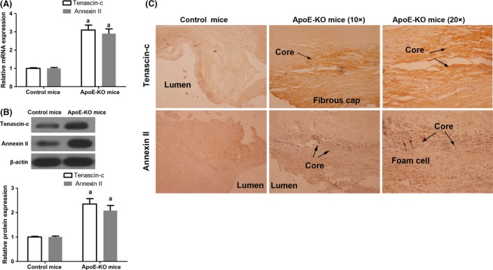
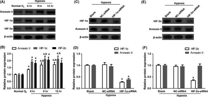
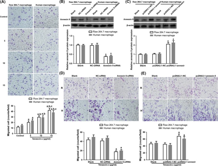
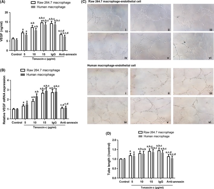
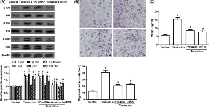
Tenascin-c is an extracellular matrix glycoprotein, the expression of which relates to the progression of atherosclerosis, myocardial infarction and heart failure. Annexin II acts as a cell surface receptor of tenascin-c. This study aimed to delineate the role of tenascin-c and annexin II in macrophages presented in atherosclerotic plaque. Animal models with atherosclerotic lesions were established using ApoE-KO mice fed with high-cholesterol diet. The expression of tenascin-c and annexin II in atherosclerotic lesions was determined by qRT-PCR, Western blot and immunohistochemistry analysis. Raw 264.7 macrophages and human primary macrophages were exposed to 5, 10 and 15 μg/ml tenascin-c for 12 hrs. Cell migration as well as the proangiogenic ability of macrophages was examined. Additionally, annexin II expression was delineated in raw 264.7 macrophages under normal condition (20% O ) for 12 hrs or hypoxic condition (1% O ) for 6-12 hrs. The expression of tenascin-c and annexin II was markedly augmented in lesion aorta. Tenascin-c positively regulated macrophage migration, which was dependent on the expression of annexin II in macrophages. VEGF release from macrophages and endothelial tube induction by macrophage were boosted by tenascin-c and attenuated by annexin II blocking. Furthermore, tenascin-c activated Akt/NF-κB and ERK signalling through annexin II. Lastly, hypoxia conditioning remarkably facilitates annexin II expression in macrophages through hypoxia-inducible factor (HIF)-1α but not HIF-2α. In conclusion, tenascin-c promoted macrophage migration and VEGF expression through annexin II, the expression of which was modulated by HIF-1α.
纤连蛋白 -C 是一种细胞外基质糖蛋白,其表达与动脉粥样硬化、心肌梗死和心力衰竭的进展有关。膜联蛋白 II 作为纤连蛋白 -C 的细胞表面受体。本研究旨在描述在动脉粥样硬化斑块中巨噬细胞中纤连蛋白 -C 和膜联蛋白 II 的作用。使用高脂饮食喂养的 ApoE-KO 小鼠建立动脉粥样硬化病变动物模型。通过 qRT-PCR、Western blot 和免疫组化分析测定动脉粥样硬化病变中纤连蛋白 -C 和膜联蛋白 II 的表达。将 Raw 264.7 巨噬细胞和人原代巨噬细胞暴露于 5、10 和 15μg/ml 的纤连蛋白 -C 中 12 小时。检查巨噬细胞的迁移和促血管生成能力。此外,在正常条件(20%O )下 12 小时或缺氧条件(1%O )下 6-12 小时描绘 Raw 264.7 巨噬细胞中膜联蛋白 II 的表达。病变主动脉中纤连蛋白 -C 和膜联蛋白 II 的表达明显增加。纤连蛋白 -C 正向调节巨噬细胞迁移,这依赖于巨噬细胞中膜联蛋白 II 的表达。巨噬细胞释放的 VEGF 和内皮管诱导由巨噬细胞增强纤连蛋白 -C 和抑制由膜联蛋白 II 阻断。此外,纤连蛋白 -C 通过膜联蛋白 II 激活 Akt/NF-κB 和 ERK 信号通路。最后,缺氧条件通过缺氧诱导因子 (HIF)-1α而不是 HIF-2α显著促进巨噬细胞中膜联蛋白 II 的表达。总之,纤连蛋白 -C 通过膜联蛋白 II 促进巨噬细胞迁移和 VEGF 表达,其表达受 HIF-1α 调节。