Zhang Wen-Jun, Zhang Zhong-Lei, Chang Jun-Jie, Song Xiao-Yu
Department of Medical Ultrasound, Taihe Hospital, Hubei University of Medicine, Shiyan, Hubei, China.
Medicine (Baltimore). 2017 Sep;96(35):e7747. doi: 10.1097/MD.0000000000007747.
Absent pulmonary valve syndrome (APVS) is a rare congenital heart disease that is often associated with tetralogy of Fallot (TOF). Here, we report 2 cases of APVS associated with TOF diagnosed via fetal echocardiography and discuss their specific ultrasonographic characteristics.
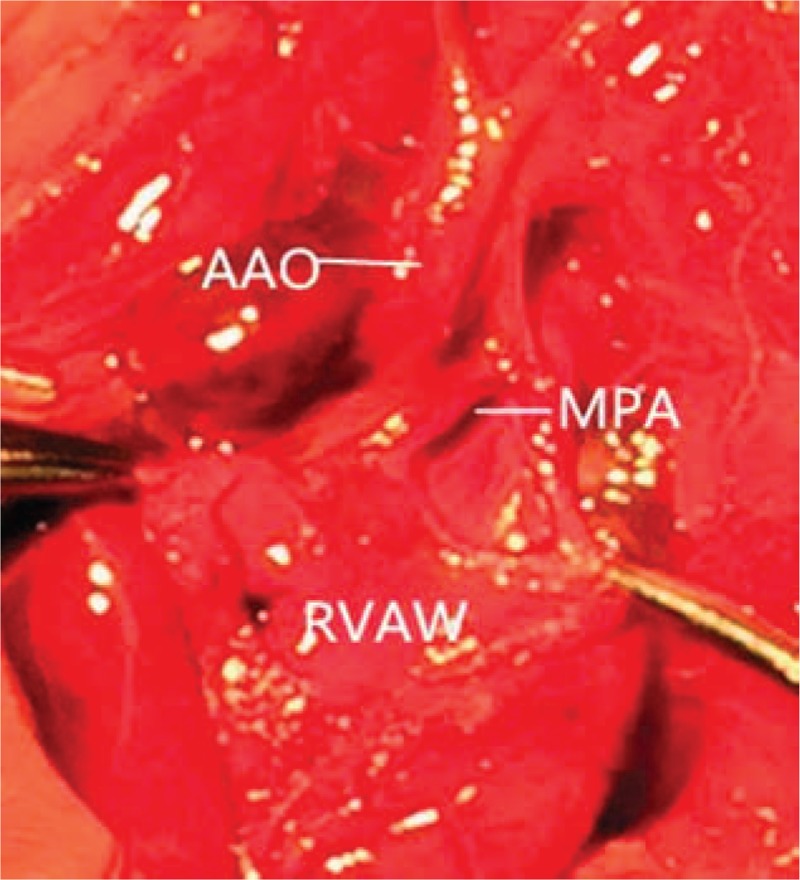
Two pregnant women with suspicion of fetal heart anomaly were referred from their local hospitals to our hospital for fetal malformation screening and detailed fetal echocardiography. Color and spectral Doppler flow imaging were utilized to evaluate the axis, size, situs, cardiac chambers, and both inflow and outflow tracts of the heart as well as the great arteries. Both cases had a severe dilatation of the pulmonary trunk and its branches and an absence or dysplasia of the pulmonary valve, which was associated with subaortic ventricular septal defect (VSD) with an overriding aorta. In addition, the fetus in case 1 showed a patent ductus arteriosus, and the fetus in case 2 showed arterial duct agenesis. Furthermore, color Doppler flow imaging showed a bi-directional multicolored flow signal in the pulmonary valve ring.
Both fetuses were diagnosed with APVS associated with TOF.
No therapeutic intervention was performed.
On the request of the pregnant women and their families, both fetuses were aborted.
Although APVS is a rare congenital heart disease and often associated with TOF, it has an overall poor prognosis. Nowadays, it can be easily diagnosed via ultrasonography because of its typical ultrasonographic features, such as aneurysmal dilatation of pulmonary artery, massive regurgitation of the pulmonary valve, VSD, and an overriding aorta. Therefore, early fetal echocardiography screening should be performed for every fetus.
肺动脉瓣缺如综合征(APVS)是一种罕见的先天性心脏病,常与法洛四联症(TOF)相关。在此,我们报告2例经胎儿超声心动图诊断为与TOF相关的APVS病例,并讨论其特定的超声特征。
两名怀疑胎儿心脏异常的孕妇从当地医院转诊至我院进行胎儿畸形筛查及详细的胎儿超声心动图检查。采用彩色和频谱多普勒血流成像评估心脏的轴线、大小、位置、心腔、流入和流出道以及大动脉。两例均有肺动脉主干及其分支的严重扩张以及肺动脉瓣缺如或发育异常,伴有主动脉下室间隔缺损(VSD)及主动脉骑跨。此外,病例1的胎儿显示动脉导管未闭,病例2的胎儿显示动脉导管缺如。此外,彩色多普勒血流成像显示肺动脉瓣环处有双向多彩血流信号。
两名胎儿均被诊断为与TOF相关的APVS。
未进行治疗干预。
应孕妇及其家属要求,两名胎儿均被引产。
尽管APVS是一种罕见的先天性心脏病且常与TOF相关,但其总体预后较差。如今,因其典型的超声特征,如肺动脉瘤样扩张、肺动脉瓣大量反流、VSD及主动脉骑跨,通过超声检查可轻易诊断。因此,应对每个胎儿进行早期胎儿超声心动图筛查。