Ermund Anna, Meiss Lauren N, Rodriguez-Pineiro Ana M, Bähr Andrea, Nilsson Harriet E, Trillo-Muyo Sergio, Ridley Caroline, Thornton David J, Wine Jeffrey J, Hebert Hans, Klymiuk Nikolai, Hansson Gunnar C
Department of Medical Biochemistry, University of Gothenburg, SE-405 30 Gothenburg, Sweden.
Institute of Molecular Animal Breeding and Biotechnology, Gene Center, Ludwig-Maximilians-University Munich, Feodor-Lynen-Straße 25, 81377 Munich, Germany.
Biochem Biophys Res Commun. 2017 Oct 21;492(3):331-337. doi: 10.1016/j.bbrc.2017.08.113. Epub 2017 Aug 30.
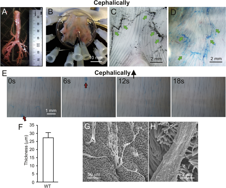
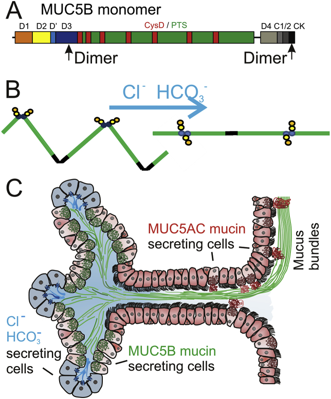
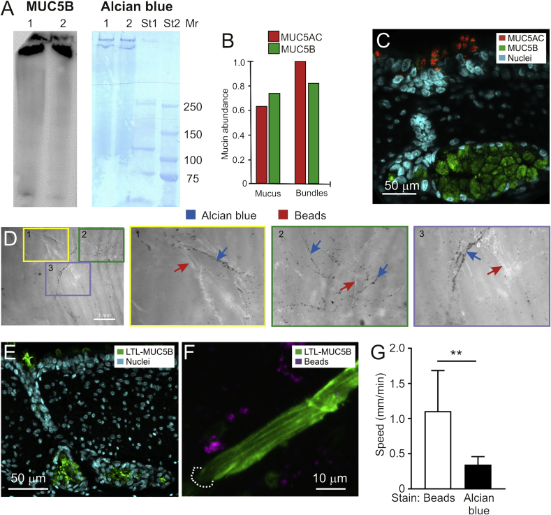
To understand the mucociliary clearance system, mucins were visualized by light, confocal and electron microscopy, and mucus was stained by Alcian blue and tracked by video microscopy on tracheal explants of newborn piglets. We observed long linear mucus bundles that appeared at the submucosal gland openings and were transported cephalically. The mucus bundles were shown by mass spectrometry and immunostaining to have a core made of MUC5B mucin and were coated with MUC5AC mucin produced by surface goblet cells. The transport speed of the bundles was slower than the airway surface liquid flow. We suggest that the goblet cell MUC5AC mucin anchors the mucus bundles and thus controls their transport. Normal clearance of the respiratory tree of pigs and humans, both rich in submucosal glands, is performed by thick and long mucus bundles.
为了解黏液纤毛清除系统,利用光学显微镜、共聚焦显微镜和电子显微镜观察了黏蛋白,并采用阿尔辛蓝对新生仔猪气管外植体上的黏液进行染色,通过视频显微镜进行追踪。我们观察到长的线性黏液束出现在黏膜下腺开口处,并向头部方向运输。通过质谱分析和免疫染色表明,黏液束的核心由MUC5B黏蛋白构成,表面杯状细胞产生的MUC5AC黏蛋白包裹在其外。黏液束的运输速度比气道表面液体流动速度慢。我们认为,杯状细胞的MUC5AC黏蛋白固定了黏液束,从而控制其运输。猪和人的呼吸道树都富含黏膜下腺,其正常清除功能是通过粗大且长的黏液束来实现的。