Härtig Wolfgang, Mages Bianca, Aleithe Susanne, Nitzsche Björn, Altmann Stephan, Barthel Henryk, Krueger Martin, Michalski Dominik
Department of Pathophysiology of Neuroglia, Paul Flechsig Institute for Brain Research, University of LeipzigLeipzig, Germany.
Department of Neurology, University of LeipzigLeipzig, Germany.
Front Integr Neurosci. 2017 Aug 15;11:15. doi: 10.3389/fnint.2017.00015. eCollection 2017.
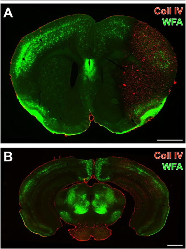
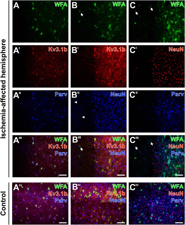
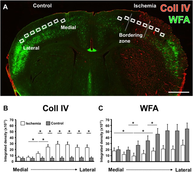
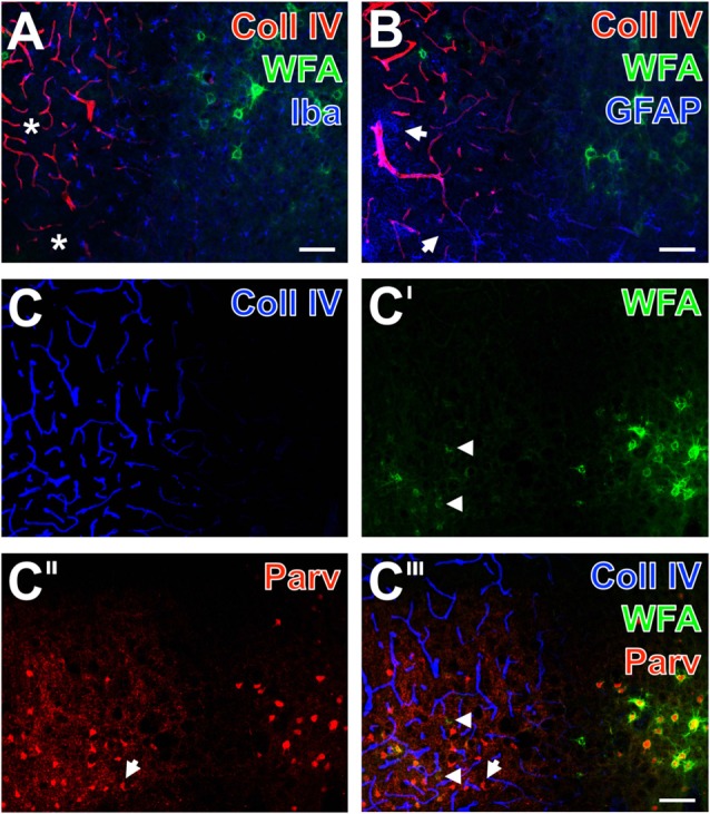
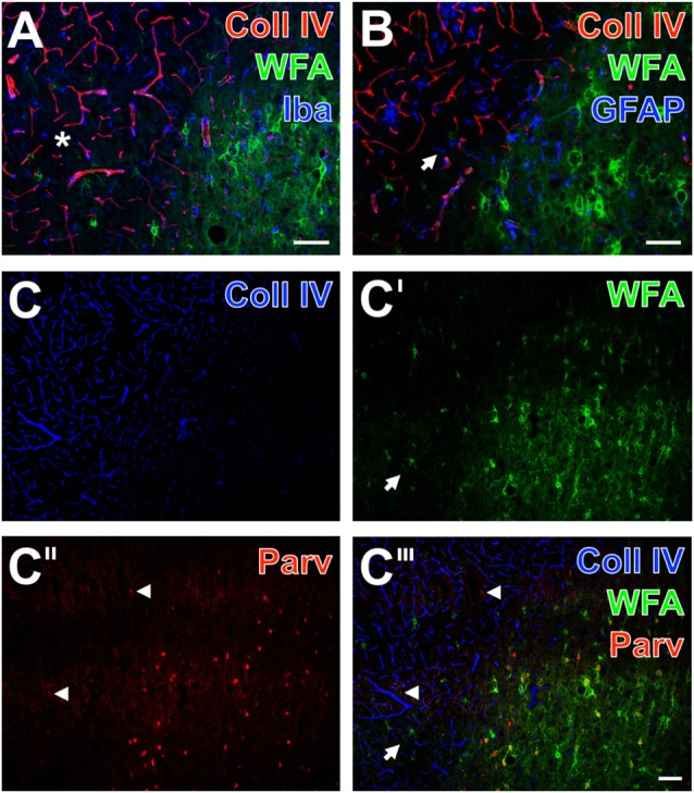
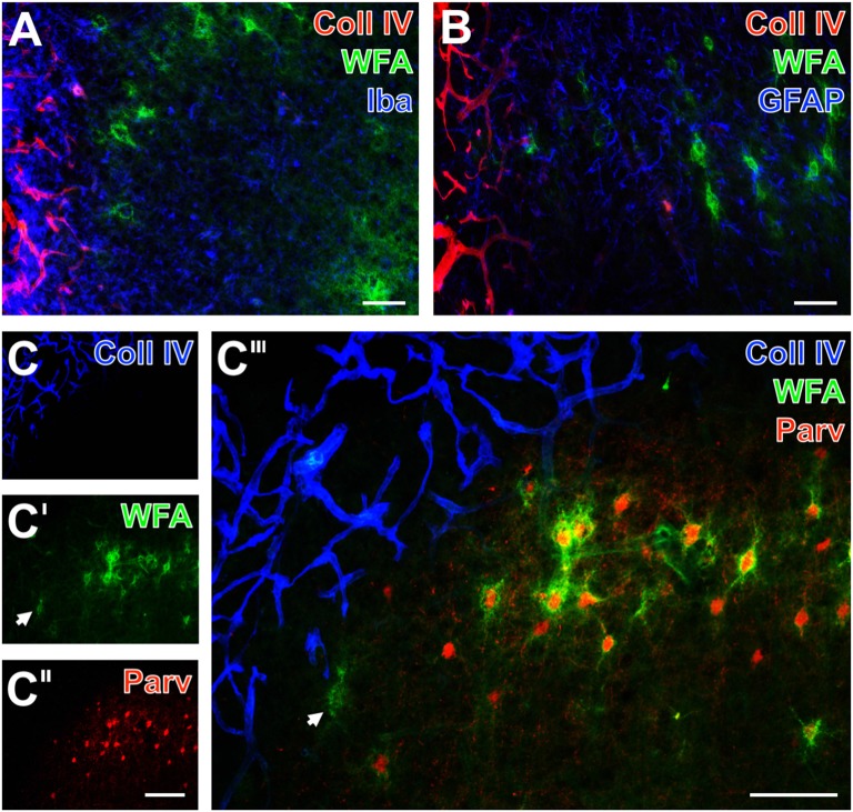
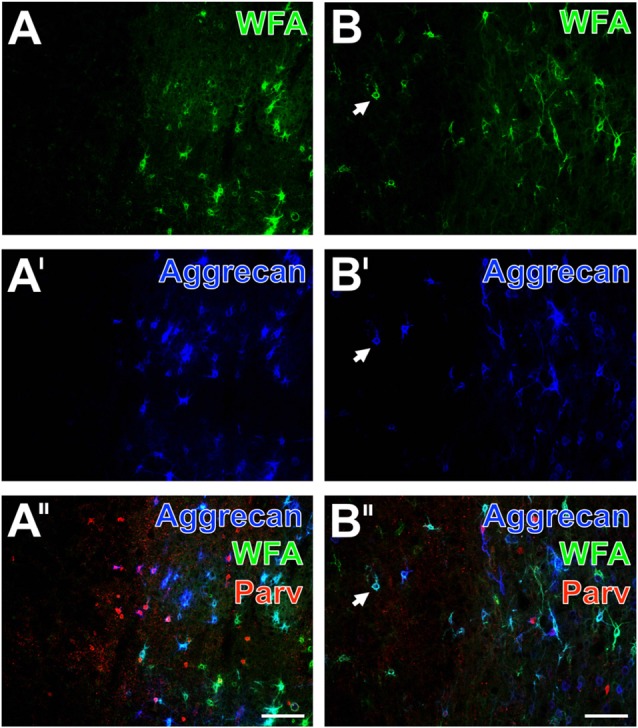
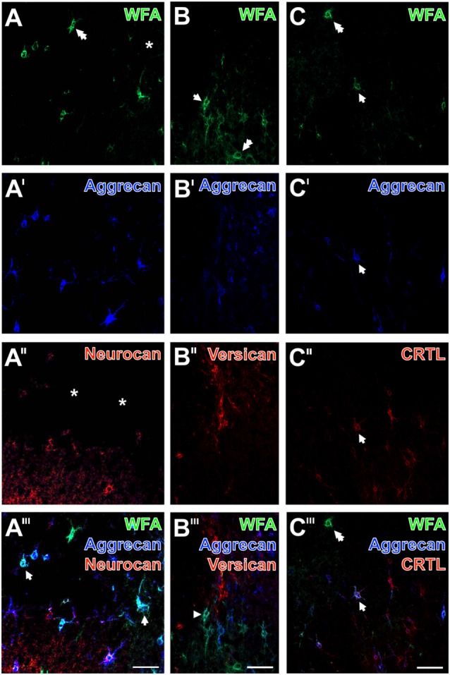
As part of the extracellular matrix (ECM), perineuronal nets (PNs) are polyanionic, chondroitin sulfate proteoglycan (CSPG)-rich coatings of certain neurons, known to be affected in various neural diseases. Although these structures are considered as important parts of the neurovascular unit (NVU), their role during evolution of acute ischemic stroke and subsequent tissue damage is poorly understood and only a few preclinical studies analyzed PNs after acute ischemic stroke. By employing three models of experimental focal cerebral ischemia, this study was focused on histopathological alterations of PNs and concomitant vascular, glial and neuronal changes according to the NVU concept. We analyzed brain tissues obtained 1 day after ischemia onset from: (a) mice after filament-based permanent middle cerebral artery occlusion (pMCAO); (b) rats subjected to thromboembolic MACO; and (c) sheep at 14 days after electrosurgically induced focal cerebral ischemia. Multiple fluorescence labeling was applied to explore simultaneous alterations of NVU and ECM. Serial mouse sections labeled with the net marker agglutinin (WFA) displayed largely decomposed and nearly erased PNs in infarcted neocortical areas that were demarcated by up-regulated immunoreactivity for vascular collagen IV (Coll IV). Subsequent semi-quantitative analyses in mice confirmed significantly decreased WFA-staining along the ischemic border zone and a relative decrease in the directly ischemia-affected neocortex. Triple fluorescence labeling throughout the three animal models revealed up-regulated Coll IV and decomposed PNs accompanied by activated astroglia and altered immunoreactivity for parvalbumin, a calcium-binding protein in fast-firing GABAergic neurons which are predominantly surrounded by neocortical PNs. Furthermore, ischemic neocortical areas in rodents simultaneously displayed less intense staining of WFA, aggrecan, the net components neurocan, versican and the cartilage link protein (CRTL) as well as markers in net-bearing neurons such as the potassium channel subunit Kv3.1b and neuronal nuclei (NeuN). In summary, theconsistent observations based on three different stroke models confirmed that PNs are highly sensitive constituents of the NVU along with impaired associated GABAergic neurons. These results suggest that PNs could be promising targets of future stroke treatment, and further studies should address their reorganization and plasticity in both stabilizing the acute stroke as well as supportive effects during the chronic phase of stroke.
作为细胞外基质(ECM)的一部分,神经元周围网(PNs)是某些神经元的富含硫酸软骨素蛋白聚糖(CSPG)的聚阴离子涂层,已知在各种神经疾病中会受到影响。尽管这些结构被认为是神经血管单元(NVU)的重要组成部分,但它们在急性缺血性中风演变及随后组织损伤过程中的作用仍知之甚少,仅有少数临床前研究分析了急性缺血性中风后的PNs。通过采用三种实验性局灶性脑缺血模型,本研究聚焦于根据NVU概念分析PNs的组织病理学改变以及伴随的血管、神经胶质和神经元变化。我们分析了缺血发作后1天获得的脑组织,这些脑组织来自:(a)基于线栓法的永久性大脑中动脉闭塞(pMCAO)后的小鼠;(b)接受血栓栓塞性大脑中动脉闭塞(MACO)的大鼠;以及(c)电外科诱导局灶性脑缺血14天后的绵羊。应用多重荧光标记来探究NVU和ECM的同时改变。用网状标记物荆豆凝集素(WFA)标记的小鼠连续切片显示,梗死的新皮质区域中PNs大多分解且几乎消失,这些区域由血管胶原IV(Coll IV)免疫反应性上调所界定。随后在小鼠中的半定量分析证实,沿缺血边界区WFA染色显著减少,而直接受缺血影响的新皮质中相对减少。在所有三种动物模型中进行的三重荧光标记显示,Coll IV上调且PNs分解,同时伴有星形胶质细胞活化以及小白蛋白免疫反应性改变,小白蛋白是快速放电的GABA能神经元中的一种钙结合蛋白,主要被新皮质PNs所包围。此外,啮齿动物的缺血新皮质区域同时显示WFA、聚集蛋白聚糖、网状成分神经蛋白聚糖、多功能蛋白聚糖和软骨连接蛋白(CRTL)以及网状神经元中的标记物如钾通道亚基Kv3.1b和神经元细胞核(NeuN)的染色强度降低。总之,基于三种不同中风模型的一致观察结果证实,PNs是NVU中高度敏感的成分,同时相关的GABA能神经元受损。这些结果表明,PNs可能是未来中风治疗的有希望的靶点,进一步的研究应探讨它们在稳定急性中风以及中风慢性期的支持作用方面的重组和可塑性。