Lakpour Mohammad Reza, Aghajanpour Samaneh, Koruji Morteza, Shahverdi Abdolhossein, Sadighi-Gilani Mohammad Ali, Sabbaghian Marjan, Aflatoonian Reza, Rajabian-Naghandar Majid
Department of Andrology, Reproductive Biomedicine Research Center, Royan Institute for Reproductive Biomedicine, ACECR, Tehran, Iran.
Biology Department, Payam Noor University, Tehran, Iran.
J Reprod Infertil. 2017 Apr-Jun;18(2):213-217.
The sertoli cells in the testis create unique and safe environment to protect seminiferous tubules from auto antigens and invading pathogens. These cells produce the survival factor of the blood-testis barrier and produce special materials such as androgen binding proteins and contribute to the coordinated action of spermatogenesis. Given that the sertoli cells play an essential role in spermatogenesis and the lack of these cells leads to the disruption of spermatogenesis, it is necessary to investigate the behavior and performance of these cells. To achieve this goal, the cells must first be extracted. The aim of this study was to develop a procedure to isolate, culture, and characterize human sertoli cells.
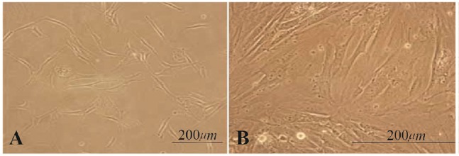
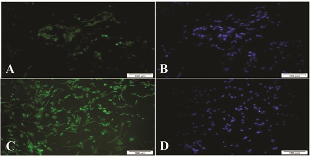
In order to isolate the sertoli cells of azoospermia patients who underwent (testicular sperm extraction) TESE surgery, washing up and multi_stage enzyme digestion of single cells, culture on petri dishes impregnated with datura stramonium lectin agglutinin (DSA) were done and then the cells were passaged for several times and isolated. For more purification, flow cytometry method with FSH receptor antibody was used. Immunocytochemistry assays and Elisa test for identification of these cells were employed.
The purification method resulted in more than 97% purity. The nature of sertoli cells was confirmed by morphology evaluation, detecting anti-mullerian hormone in sertoli cell culture media and the presence of FSH receptor on sertoli cells.
This study introduced and applied a method to isolate, culture, and purify human sertoli cells with high purity which made possible further researches on these cells.
睾丸中的支持细胞营造了独特且安全的环境,以保护生精小管免受自身抗原和入侵病原体的侵害。这些细胞产生血睾屏障的存活因子,并产生诸如雄激素结合蛋白等特殊物质,有助于精子发生的协同作用。鉴于支持细胞在精子发生中起着至关重要的作用,且这些细胞的缺失会导致精子发生中断,因此有必要研究这些细胞的行为和性能。为实现这一目标,必须首先提取这些细胞。本研究的目的是开发一种分离、培养和鉴定人支持细胞的方法。
为了分离接受睾丸精子提取(TESE)手术的无精子症患者的支持细胞,进行了单细胞的冲洗和多阶段酶消化,在浸有曼陀罗凝集素(DSA)的培养皿上培养,然后对细胞进行多次传代并分离。为了进一步纯化,使用了抗促卵泡激素(FSH)受体抗体的流式细胞术方法。采用免疫细胞化学分析和酶联免疫吸附测定(ELISA)来鉴定这些细胞。
纯化方法使纯度超过97%。通过形态学评估、检测支持细胞培养基中的抗苗勒管激素以及支持细胞上FSH受体的存在,证实了支持细胞的性质。
本研究介绍并应用了一种分离、培养和纯化高纯度人支持细胞的方法,这使得对这些细胞的进一步研究成为可能。