Masum Md Abdul, Ichii Osamu, Elewa Yaser Hosny Ali, Nakamura Teppei, Kon Yasuhiro
Laboratory of Anatomy, Graduate School of Veterinary Medicine, Hokkaido University, Kita 18, Nishi 9, Kita-ku, Sapporo, Japan.
Department of Histology, Faculty of Veterinary Medicine, Zagazig University, Zagazig, Egypt.
BMC Nephrol. 2017 Sep 4;18(1):280. doi: 10.1186/s12882-017-0694-3.
The renal vasculature plays important roles in both homeostasis and pathology. In this study, we examined pathological changes in the renal microvascular in mouse models of kidney diseases.
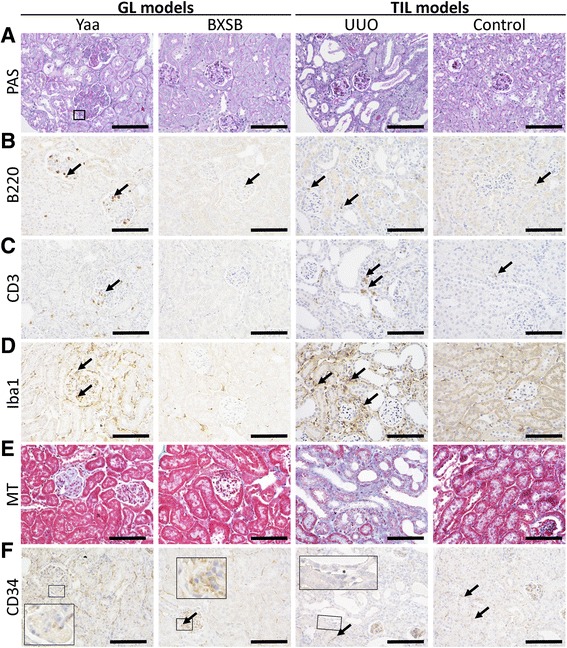
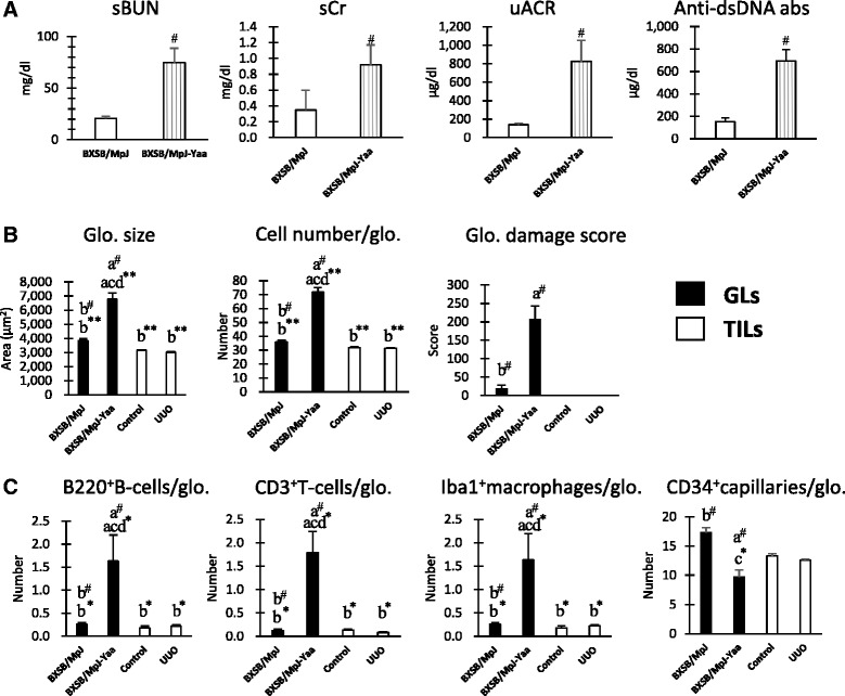
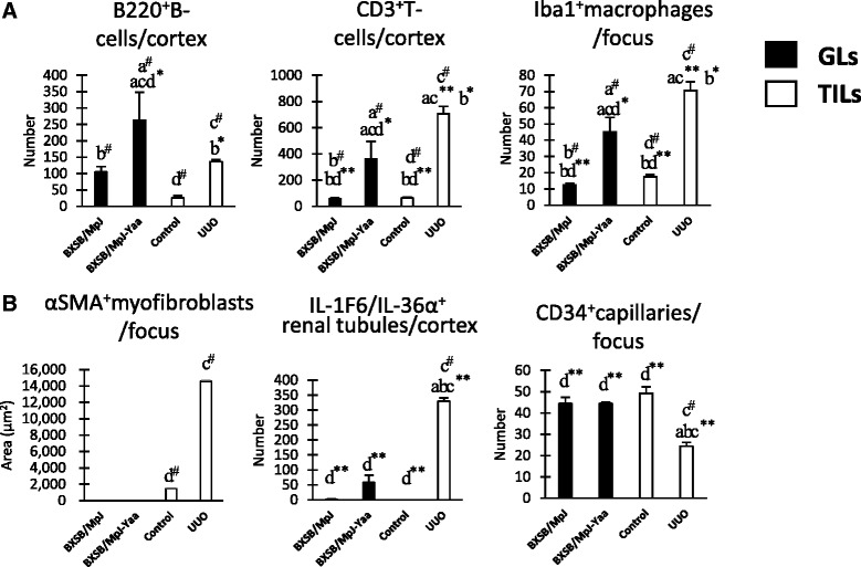
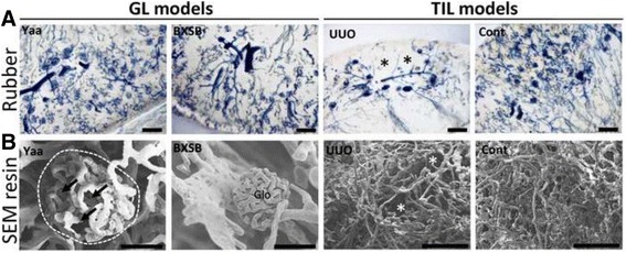
Glomerular lesions (GLs) in autoimmune disease-prone male BXSB/MpJ-Yaa (Yaa) mice and tubulointerstitial lesions (TILs) in male C57BL/6 mice subjected to unilateral ureteral obstruction (UUO) for 7 days were studied. Collected kidneys were examined using histopathological techniques. A nonparametric Mann-Whitney U test (P < 0.05) was performed to compare healthy controls and the experimental mice. The Kruskal-Wallis test was used to compare three or more groups, and multiple comparisons were performed using Scheffe's method when significant differences were observed (P < 0.05).
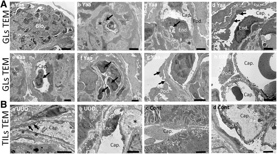
Yaa mice developed severe autoimmune glomerulonephritis, and the number of CD34 glomerular capillaries decreased significantly in GLs compared to that in control mice. However, UUO-treated mice showed severe TILs only, and CD34 tubulointerstitial capillaries were decreased significantly in TILs with the progression of tubulointerstitial fibrosis compared to those in untreated control kidneys. Infiltrations of B-cells, T-cells, and macrophages increased significantly in the respective lesions of both disease models (P < 0.05). In observations of vascular corrosion casts by scanning electron microscopy and of microfil rubber-perfused thick kidney sections by fluorescence microscopy, segmental absences of capillaries were observed in the GLs and TILs of Yaa and UUO-treated mice, respectively. Further, transmission electron microscopy revealed capillary endothelial injury in the respective lesions of both models. The numbers of CD34 glomerular and tubulointerstitial capillaries were negatively correlated with all examined parameters in GLs (P < 0.05) and TILs (P < 0.01), respectively.
From the analysis of mouse models, we identified inverse pathological correlations between the number of local capillaries in GLs and TILs and the severity of kidney diseases.
肾血管系统在体内平衡和病理过程中均发挥着重要作用。在本研究中,我们检测了肾脏疾病小鼠模型肾微血管的病理变化。
研究了自身免疫病易感雄性BXSB/MpJ-Yaa(Yaa)小鼠的肾小球病变(GLs)以及单侧输尿管梗阻(UUO)7天的雄性C57BL/6小鼠的肾小管间质病变(TILs)。使用组织病理学技术检查收集的肾脏。采用非参数曼-惠特尼U检验(P < 0.05)比较健康对照小鼠和实验小鼠。使用Kruskal-Wallis检验比较三组或更多组,当观察到显著差异(P < 0.05)时,使用Scheffe方法进行多重比较。
Yaa小鼠发生了严重的自身免疫性肾小球肾炎,与对照小鼠相比,GLs中CD34肾小球毛细血管数量显著减少。然而,UUO处理的小鼠仅表现出严重的TILs,与未处理的对照肾脏相比,随着肾小管间质纤维化的进展,TILs中CD34肾小管间质毛细血管显著减少。在两种疾病模型的各自病变中,B细胞、T细胞和巨噬细胞的浸润均显著增加(P < 0.05)。通过扫描电子显微镜观察血管铸型以及通过荧光显微镜观察微丝橡胶灌注的肾脏厚切片时,分别在Yaa小鼠和UUO处理小鼠的GLs和TILs中观察到毛细血管节段性缺失。此外,透射电子显微镜显示两种模型各自病变中的毛细血管内皮损伤。GLs(P < 0.05)和TILs(P < 0.01)中CD34肾小球和肾小管间质毛细血管数量分别与所有检测参数呈负相关。
通过对小鼠模型的分析,我们确定了GLs和TILs中局部毛细血管数量与肾脏疾病严重程度之间的反向病理相关性。