Benedetto María M, Guido Mario E, Contin María A
Facultad de Ciencias Químicas, Departamento de Química Biológica "Dr. Ranwel Caputto", Universidad Nacional de Córdoba, Córdoba, Argentina.
Centro de Investigaciones en Química Biológica de Córdoba (CIQUIBIC), CONICET, Universidad Nacional de Córdoba, Córdoba, Argentina.
Front Neurol. 2017 Aug 21;8:417. doi: 10.3389/fneur.2017.00417. eCollection 2017.
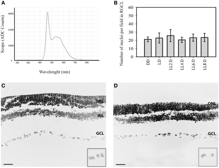
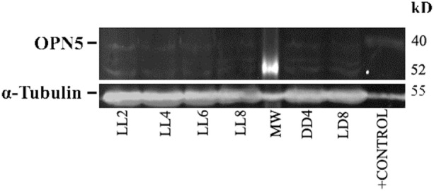
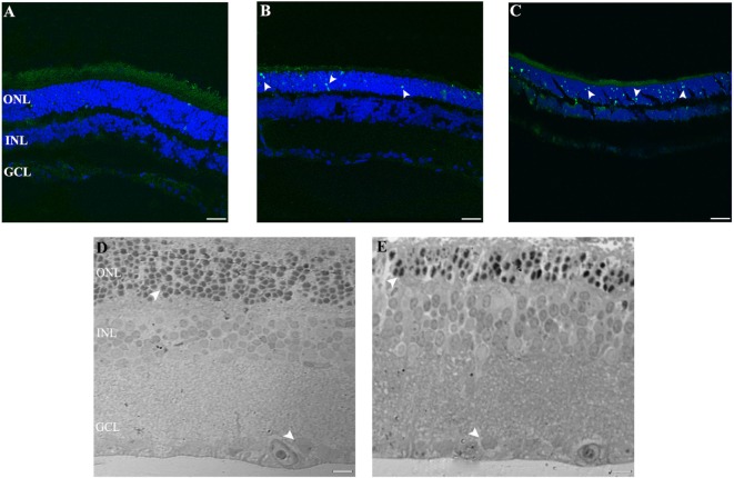
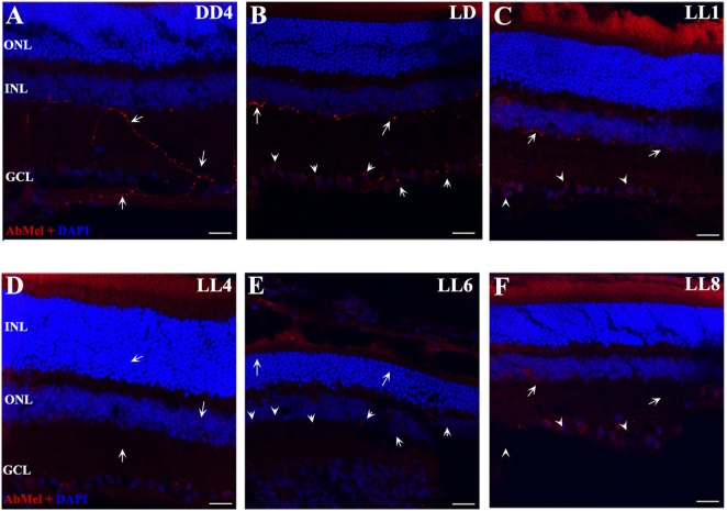
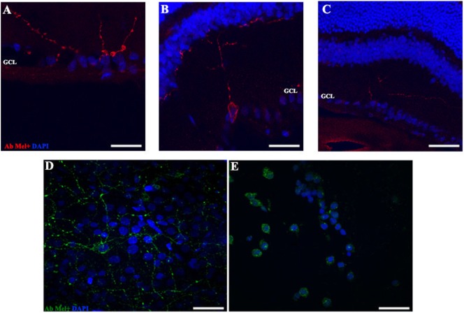
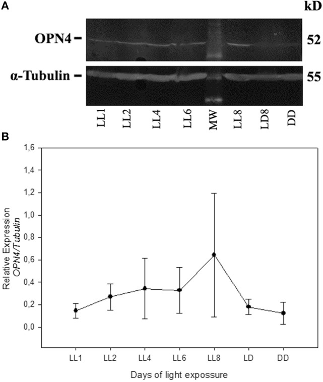
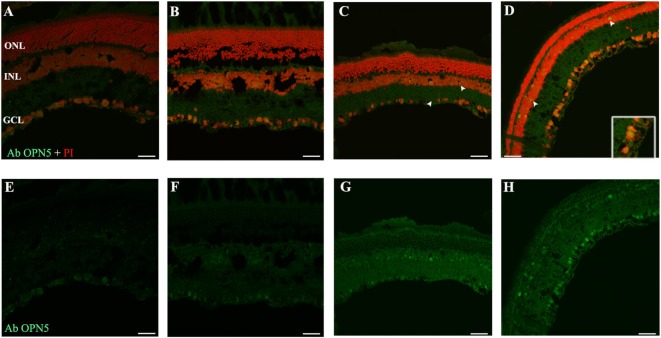
The retina is part of the central nervous system specially adapted to capture light photons and transmit this information to the brain through photosensitive retinal cells involved in visual and non-visual activities. However, excessive light exposure may accelerate genetic retinal diseases or induce photoreceptor cell (PRC) death, finally leading to retinal degeneration (RD). Light pollution (LP) caused by the characteristic use of artificial light in modern day life may accelerate degenerative diseases or promote RD and circadian desynchrony. We have developed a working model to study RD mechanisms in a low light environment using light-emitting diode (LED) sources, at constant or long exposure times under LP conditions. The mechanism of PRC death is still not fully understood. Our main goal is to study the biochemical mechanisms of RD. We have previously demonstrated that constant light (LL) exposure to white LED produces a significant reduction in the outer nuclear layer (ONL) by classical PRC death after 7 days of LL exposure. The PRCs showed TUNEL-positive labeling and a caspase-3-independent mechanism of cell death. Here, we investigate whether constant LED exposure affects the inner-retinal organization and structure, cell survival and the expression of photopigments; in particular we look into whether constant LED exposure causes the death of retinal ganglion cells (RGCs), of intrinsically photosensitive RGCs (ipRGCs), or of other inner-retinal cells. rats exposed to 200 lx of LED for 2 to 8 days (LL 2 and LL 8) were processed for histological and protein. The results show no differences in the number of nucleus or TUNEL positive RGCs nor inner structural damage in any of LL groups studied, indicating that LL exposure affects ONL but does not produce RGC death. However, the photopigments melanopsin (OPN4) and neuropsin (OPN5) expressed in the inner retina were seen to modify their localization and expression during LL exposure. Our findings suggest that constant light during several days produces retinal remodeling and ONL cell death as well as significant changes in opsin expression in the inner nuclear layer.
视网膜是中枢神经系统的一部分,它经过特殊适配,能够捕获光光子,并通过参与视觉和非视觉活动的光敏视网膜细胞将这些信息传递给大脑。然而,过度暴露于光线下可能会加速遗传性视网膜疾病的发展或导致光感受器细胞(PRC)死亡,最终导致视网膜变性(RD)。现代生活中人工光的特殊使用所造成的光污染(LP)可能会加速退行性疾病的发展或促进视网膜变性和昼夜节律失调。我们已经开发出一种工作模型,用于在低光照环境下,使用发光二极管(LED)光源,在LP条件下以恒定或长时间暴露的方式研究视网膜变性的机制。PRC死亡的机制仍未完全明确。我们的主要目标是研究视网膜变性的生化机制。我们之前已经证明,持续暴露于白色LED的恒定光(LL)下7天后,经典的PRC死亡会导致外核层(ONL)显著减少。PRC显示出TUNEL阳性标记以及一种不依赖于半胱天冬酶-3的细胞死亡机制。在这里,我们研究持续的LED暴露是否会影响视网膜内层的组织和结构、细胞存活以及光色素的表达;特别是我们要探究持续的LED暴露是否会导致视网膜神经节细胞(RGC)、内在光敏性RGC(ipRGC)或其他视网膜内层细胞死亡。将暴露于200勒克斯LED光下2至8天的大鼠(LL 2和LL 8)进行组织学和蛋白质分析。结果显示,在所研究的任何LL组中,细胞核数量或TUNEL阳性RGC数量均无差异,也没有内部结构损伤,这表明LL暴露会影响ONL,但不会导致RGC死亡。然而,在内视网膜中表达 的光色素黑视蛋白(OPN4)和神经视蛋白(OPN5)在LL暴露期间其定位和表达发生了改变。我们的研究结果表明,持续数天的光照会导致视网膜重塑和ONL细胞死亡,以及内核层视蛋白表达的显著变化。