Miyamoto Yutaka, Kanzaki Hiroyuki, Wada Satoshi, Tsuruoka Sari, Itohiya Kanako, Kumagai Kenichi, Hamada Yoshiki, Nakamura Yoshiki
Department of orthodontics, School of Dental Medicine, Tsurumi University, 2-1-3 Tsurumi, Tsurumi-ku, Yokohama 230-8501, Kanagawa Pref., Japan.
Department of Oral and Maxillofacial Surgery, School of Dental Medicine, Tsurumi University, 2-1-3 Tsurumi, Tsurumi-ku, Yokohama 230-8501, Kanagawa Pref., Japan.
Bone Rep. 2017 Jul 23;7:41-50. doi: 10.1016/j.bonr.2017.07.002. eCollection 2017 Dec.
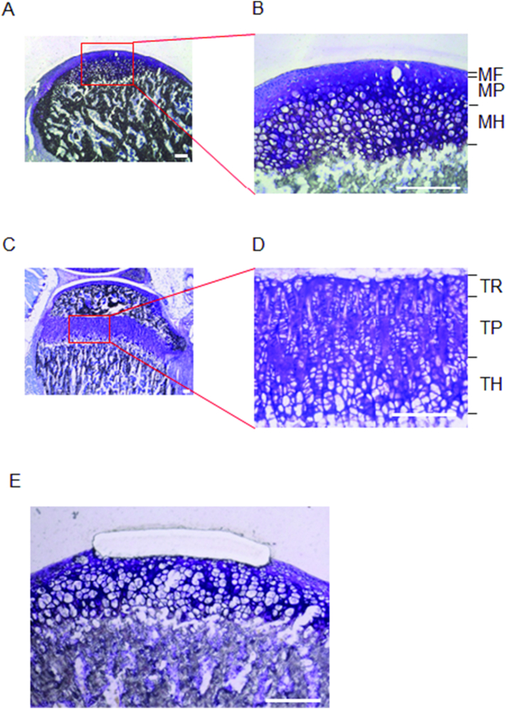
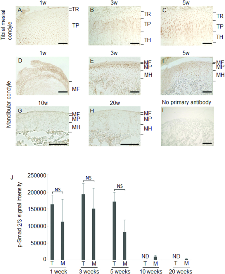
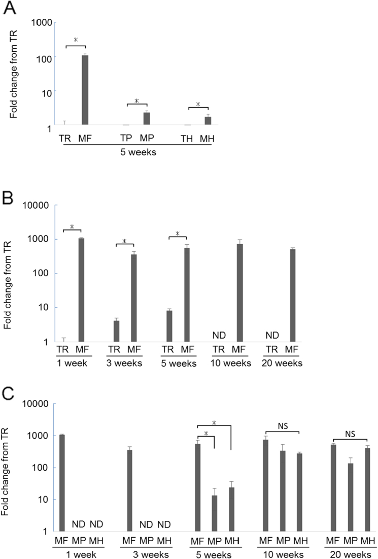
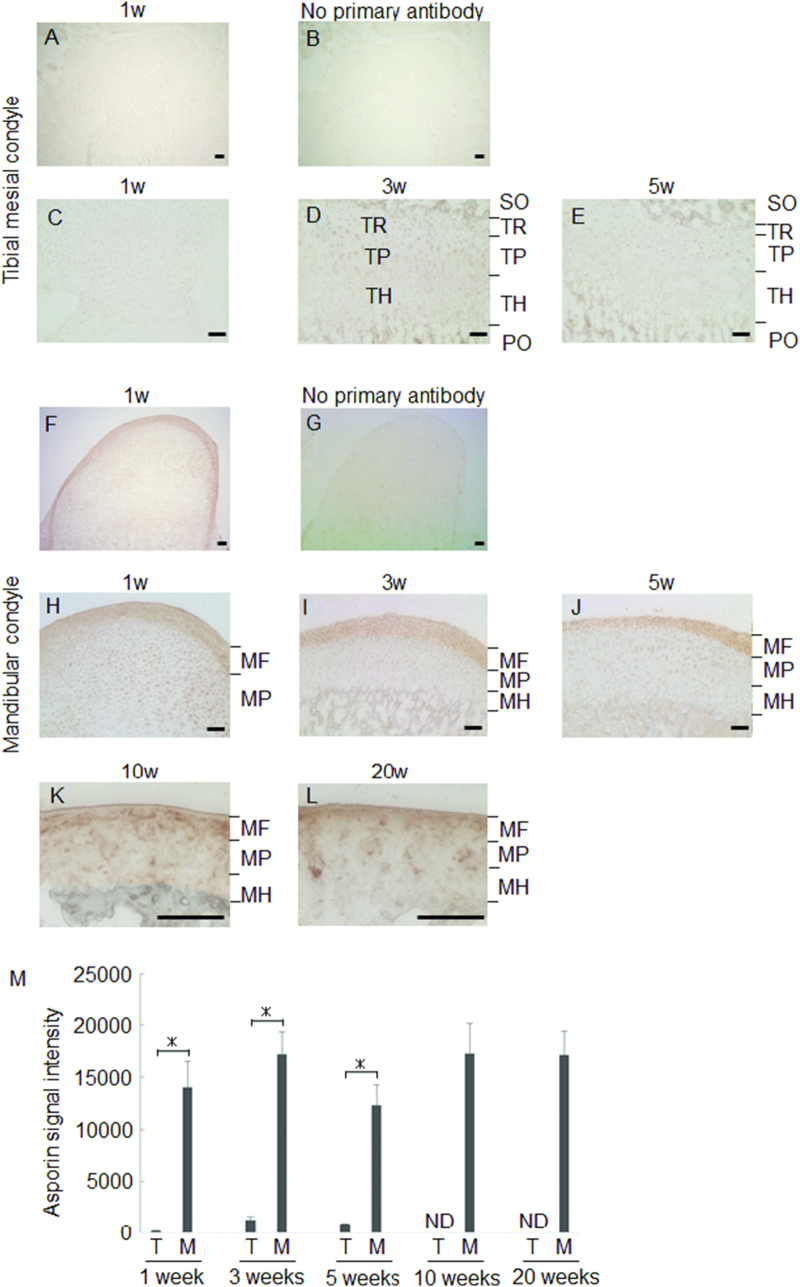
Mandibular condylar cartilage (MCC) exhibits dual roles both articular cartilage and growth center. Of many growth factors, TGF-β has been implicated in the growth of articular cartilage including MCC. Recently, Asporin, decoy to TGF-β, was discovered and it blocks TGF-β signaling. Asporin is expressed in a variety of tissues including osteoarthritic articular cartilage, though there was no report of Asporin expression in MCC. In the present study, we investigated the temporal and spatial expression of Asporin in MCC. Gene expression profile of MCC and epiphyseal cartilage in tibia of 5 weeks old ICR mice were firstly compared with microarray analysis using the laser capture microdissected samples. Variance of gene expression was further confirmed by real-time RT-PCR and immunohistochemical staining at 1,3,10, and 20 weeks old. TGF-β and its signaling molecule, phosphorylated Smad-2/3 (p-Smad2/3), were also examined by immunohistochemical staining. Microarray analysis revealed that Asporin was highly expressed in MCC. Real-time RT-PCR analysis confirmed that the fibrous layer of MCC exhibited stable higher Asporin expression at any time points as compared to epiphyseal cartilage. This was also observed in immunohistochemical staining. Deeper layer in MCC augmented Asporin expression with age. Whereas, TGF-β was stably highly observed in the layer. The fibrous layer of MCC exhibited weak staining of p-Smad2/3, though the proliferating layer of MCC was strongly stained as compared to epiphyseal cartilage of tibia at early time point. Consistent with the increase of Asporin expression in the deeper layer of MCC, the intensity of p-Smad-2/3 staining was decreased with age. In conclusion, we discovered that Asporin was stably expressed at the fibrous layer of MCC, which makes it possible to manage both articular cartilage and growth center at the same time.
下颌髁突软骨(MCC)兼具关节软骨和生长中心的双重作用。在众多生长因子中,转化生长因子-β(TGF-β)与包括MCC在内的关节软骨生长有关。最近,发现了TGF-β的诱饵蛋白——抗酒石酸酸性磷酸酶(Asporin),它可阻断TGF-β信号传导。Asporin在多种组织中表达,包括骨关节炎关节软骨,不过尚无关于其在MCC中表达的报道。在本研究中,我们调查了Asporin在MCC中的时空表达。首先,使用激光捕获显微切割样本,通过微阵列分析比较了5周龄ICR小鼠胫骨中MCC和骨骺软骨的基因表达谱。通过实时逆转录-聚合酶链反应(RT-PCR)以及1、3、10和20周龄时的免疫组织化学染色进一步证实了基因表达的差异。还通过免疫组织化学染色检测了TGF-β及其信号分子磷酸化Smad-2/3(p-Smad2/3)。微阵列分析显示,Asporin在MCC中高表达。实时RT-PCR分析证实,与骨骺软骨相比,MCC的纤维层在任何时间点均表现出稳定的更高Asporin表达。免疫组织化学染色也观察到了这一点。MCC的深层随着年龄增长Asporin表达增加。而TGF-β在该层中稳定高表达。MCC的纤维层p-Smad2/3染色较弱,不过在早期时间点,与胫骨骨骺软骨相比,MCC的增殖层染色较强。与MCC深层中Asporin表达的增加一致,p-Smad-2/3染色强度随年龄降低。总之,我们发现Asporin在MCC的纤维层中稳定表达,这使得同时管理关节软骨和生长中心成为可能。