Nokes Brandon T, Baumann Coralie P, Cummings Kristopher W, Larsen Brandon T, Cartin-Ceba Rodrigo, Swanson Karen L
The Department of Internal Medicine, Mayo Clinic, Scottsdale, AZ, United States.
The Division of Pulmonary Medicine, Mayo Clinic, Scottsdale, AZ, United States.
Respir Med Case Rep. 2017 Aug 24;22:209-211. doi: 10.1016/j.rmcr.2017.08.018. eCollection 2017.
Peripheral nerve sheath tumors (PNST) are exceedingly rare, especially outside of the posterior mediastinum. These tumors represent less than 1% of pulmonary tumors. Very few pulmonary PNSTs are ganglioneuromas. We present a case of a ganglioneuroma presenting as an endobronchial mass.
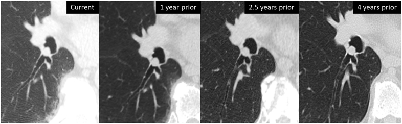
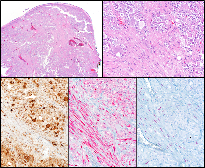
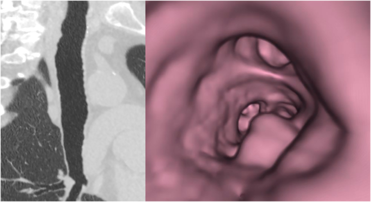
An 80 year old male was seen in pulmonary clinic for routine cancer screening. He had a 60-pack year smoking history. CT evaluation noted a 1cm right lower lobe endobronchial lesion. This lesion was present since 2012 and had slightly increased in size since that time from 8mm (Figure 1). The lesion was further assessed using virtual bronchoscopy (Figure 2). Bronchoscopy revealed an obstructing lesion, which was completely excised with the snare (Figure 3). Pathology revealed well-circumscribed tumor consisting of nests and trabeculae of round/polygonal cells with granular eosinophilic and basophilic cytoplasm. The tumor was chromogranin, synaptophysin, S-100, pancytokeratin, SOX10, and TTF-1 positive, consistent with a ganglioneuroma.
Aside from a solitary article regarding 75 patient samples (which included only one ganglioneuroma) only a small number of intrathoracic PNSTs have been reported. Only a single case report of an endobronchial ganglioneuroma has been reported. Each of these lesions were benign, and detected on routine imaging evaluations.
An intrapulmonary endobronchial location for a PNST is an exceedingly rare presentation of an already uncommon pathology.
周围神经鞘瘤(PNST)极为罕见,尤其是在后纵隔之外。这些肿瘤占肺部肿瘤的比例不到1%。肺PNST中神经节神经瘤非常少见。我们报告一例以支气管内肿块形式出现的神经节神经瘤病例。
一名80岁男性因常规癌症筛查就诊于肺部诊所。他有60年的吸烟史。CT评估发现右肺下叶有一个1厘米的支气管内病变。该病变自2012年就已存在,自那时起大小略有增加,从8毫米增大到1厘米(图1)。使用虚拟支气管镜对该病变进行了进一步评估(图2)。支气管镜检查发现一个阻塞性病变,用圈套器将其完整切除(图3)。病理显示肿瘤边界清晰,由圆形/多边形细胞巢和小梁组成,细胞质呈嗜酸性和嗜碱性颗粒状。肿瘤嗜铬粒蛋白、突触素、S-100、全细胞角蛋白、SOX10和TTF-1均呈阳性,符合神经节神经瘤的表现。
除了一篇关于75例患者样本的文章(其中仅包括一例神经节神经瘤)外,仅报道了少数胸内PNST病例。仅报道了一例支气管内神经节神经瘤的病例报告。这些病变均为良性,在常规影像学评估中被发现。
PNST位于肺内支气管内是一种本就不常见的病理情况的极为罕见的表现形式。