• 文献检索
  • 文档翻译
  • 深度研究
  • 学术资讯
  • Suppr Zotero 插件Zotero 插件
  • 邀请有礼
  • 套餐&价格
  • 历史记录
应用&插件
Suppr Zotero 插件Zotero 插件浏览器插件Mac 客户端Windows 客户端微信小程序
定价
高级版会员购买积分包购买API积分包
服务
文献检索文档翻译深度研究API 文档MCP 服务
关于我们
关于 Suppr公司介绍联系我们用户协议隐私条款
关注我们

Suppr 超能文献

核心技术专利:CN118964589B侵权必究
粤ICP备2023148730 号-1Suppr @ 2026

文献检索

告别复杂PubMed语法,用中文像聊天一样搜索,搜遍4000万医学文献。AI智能推荐,让科研检索更轻松。

立即免费搜索

文件翻译

保留排版,准确专业,支持PDF/Word/PPT等文件格式,支持 12+语言互译。

免费翻译文档

深度研究

AI帮你快速写综述,25分钟生成高质量综述,智能提取关键信息,辅助科研写作。

立即免费体验

基于断层超声成像和三维磁共振成像的联合模型诊断产后肛提肌撕裂。

Combination of tomographic ultrasound imaging and three-dimensional magnetic resonance imaging-based model to diagnose postpartum levator avulsion.

机构信息

Department of Ultrasound in Medicine, Shanghai Jiao Tong University Affiliated Sixth People's Hospital, Shanghai, China.

Shanghai Institute of Ultrasound in Medicine, Shanghai, China.

出版信息

Sci Rep. 2017 Sep 11;7(1):11235. doi: 10.1038/s41598-017-08201-9.

DOI:10.1038/s41598-017-08201-9
PMID:28894152
原文链接:https://pmc.ncbi.nlm.nih.gov/articles/PMC5593956/
Abstract

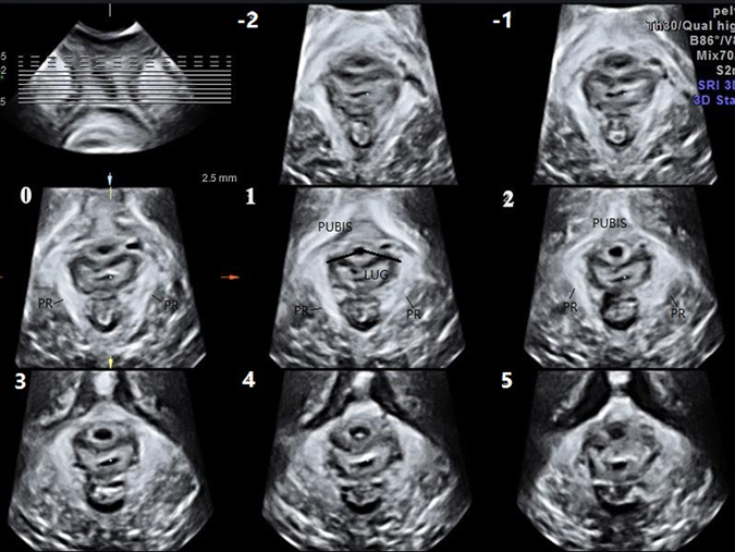
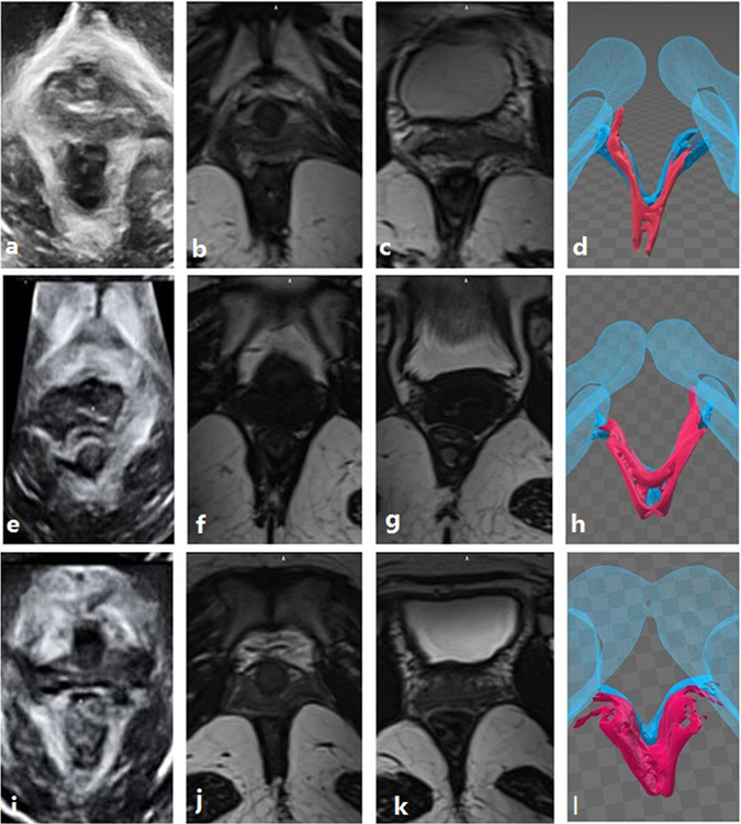
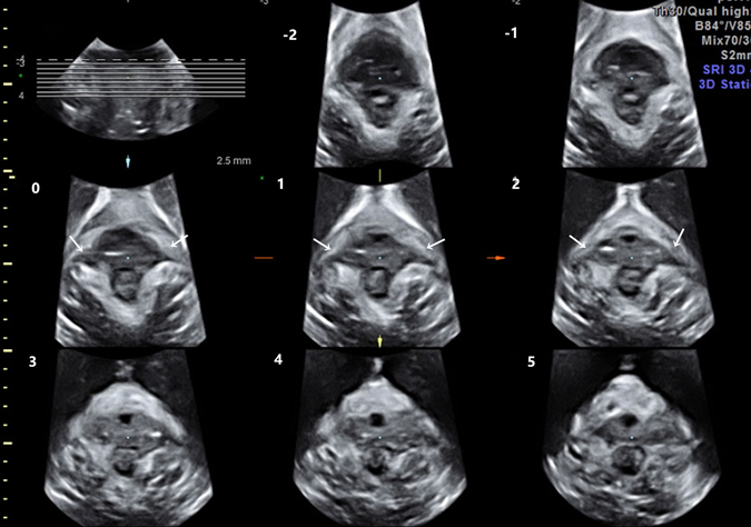
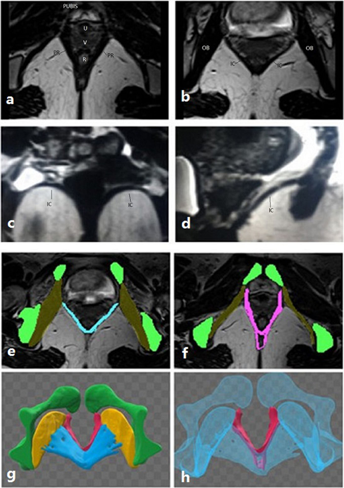
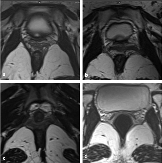
Vaginal delivery may cause levator avulsion, which may increase the risk of pelvic floor dysfunction (PFD). To explore the morphological changes of the levator ani muscle (including the puborectalis and iliococcygeus) and levator avulsion after vaginal delivery, translabial tomographic ultrasound imaging (TUI) was used to examine 80 women 45-60 days after their vaginal delivery. Subsequently, magnetic resonance imaging (MRI) was performed if at least one-sided puborectalis avulsion was found on TUI. The incidence of puborectalis avulsion in these postpartum women was 13.75% in this study. Both MRI and TUI can detect puborectalis avulsion well, and their results have good consistency. Iliococcygeus muscle injury is difficult to detect using TUI. However, MRI is a good way to observe the morphological changes of the iliococcygeus, which may also be damaged during vaginal delivery. Interestingly, our study reveals that iliococcygeus muscle injury is often associated with severe puborectalis muscle tear.

摘要

阴道分娩可能导致会阴撕裂,这可能增加盆底功能障碍(PFD)的风险。为了探讨阴道分娩后肛提肌(包括耻骨直肠肌和髂尾肌)和会阴撕裂的形态变化,本研究使用经阴道断层超声成像(TUI)检查了 80 名阴道分娩后 45-60 天的女性。随后,如果在 TUI 上发现至少一侧耻骨直肠肌撕裂,则进行磁共振成像(MRI)检查。在这些产后女性中,耻骨直肠肌撕裂的发生率为 13.75%。MRI 和 TUI 均可很好地检测耻骨直肠肌撕裂,且两者结果具有良好的一致性。TUI 难以检测髂尾肌损伤。然而,MRI 是观察髂尾肌形态变化的良好方法,其在阴道分娩过程中也可能受损。有趣的是,我们的研究表明,髂尾肌损伤常与严重的耻骨直肠肌撕裂有关。

https://cdn.ncbi.nlm.nih.gov/pmc/blobs/8f2f/5593956/50ccc59d7604/41598_2017_8201_Fig5_HTML.jpg
https://cdn.ncbi.nlm.nih.gov/pmc/blobs/8f2f/5593956/4450ec75a18a/41598_2017_8201_Fig1_HTML.jpg
https://cdn.ncbi.nlm.nih.gov/pmc/blobs/8f2f/5593956/7bd3956daf23/41598_2017_8201_Fig2_HTML.jpg
https://cdn.ncbi.nlm.nih.gov/pmc/blobs/8f2f/5593956/ab75aa35f3e6/41598_2017_8201_Fig3_HTML.jpg
https://cdn.ncbi.nlm.nih.gov/pmc/blobs/8f2f/5593956/8e1938c54fb0/41598_2017_8201_Fig4_HTML.jpg
https://cdn.ncbi.nlm.nih.gov/pmc/blobs/8f2f/5593956/50ccc59d7604/41598_2017_8201_Fig5_HTML.jpg

相似文献

1
Combination of tomographic ultrasound imaging and three-dimensional magnetic resonance imaging-based model to diagnose postpartum levator avulsion.基于断层超声成像和三维磁共振成像的联合模型诊断产后肛提肌撕裂。
Sci Rep. 2017 Sep 11;7(1):11235. doi: 10.1038/s41598-017-08201-9.
2
Changes in global strain of puborectalis muscle during pregnancy and postpartum.妊娠及产后耻骨直肠肌肌全球应变的变化。
Ultrasound Obstet Gynecol. 2018 Apr;51(4):537-542. doi: 10.1002/uog.17488.
3
Longitudinal follow-up of levator ani muscle avulsion: does a second delivery affect it?肛提肌撕裂的纵向随访:再次分娩会对其产生影响吗?
Ultrasound Obstet Gynecol. 2017 Jul;50(1):110-115. doi: 10.1002/uog.16009. Epub 2017 Jun 5.
4
Perineal and vaginal tears are clinical markers for occult levator ani trauma: a retrospective observational study.会阴及阴道撕裂是肛提肌隐匿性创伤的临床标志:一项回顾性观察研究。
Ultrasound Obstet Gynecol. 2016 Feb;47(2):224-7. doi: 10.1002/uog.14856. Epub 2016 Jan 13.
5
Pelvic floor morphometry and function in women with and without puborectalis avulsion in the early postpartum period.产后早期伴有和不伴有耻骨直肠肌撕裂的女性盆底形态和功能。
Am J Obstet Gynecol. 2017 Mar;216(3):274.e1-274.e8. doi: 10.1016/j.ajog.2016.11.1049. Epub 2016 Dec 9.
6
Injury-associated levator ani muscle and anal sphincter ooedema following vaginal birth: a secondary analysis of the EMRLD study.阴道分娩后与损伤相关的肛提肌和肛门括约肌水肿:EMRLD 研究的二次分析。
BJOG. 2021 Nov;128(12):2046-2053. doi: 10.1111/1471-0528.16760. Epub 2021 Jun 7.
7
Impact of mode of delivery on levator morphology: a prospective observational study with three-dimensional ultrasound early in the postpartum period.分娩方式对肛提肌形态的影响:产后早期三维超声前瞻性观察研究。
BJOG. 2012 Jan;119(1):51-60. doi: 10.1111/j.1471-0528.2011.03152.x. Epub 2011 Oct 10.
8
Vacuum-assisted vaginal delivery and levator ani avulsion in primiparous women.初产妇的真空辅助阴道分娩与肛提肌撕裂
J Matern Fetal Neonatal Med. 2016;29(16):2715-8. doi: 10.3109/14767058.2015.1102223. Epub 2015 Nov 9.
9
Levator avulsion using a tomographic ultrasound and magnetic resonance-based model.基于断层超声和磁共振的提肌撕脱术。
Am J Obstet Gynecol. 2011 Sep;205(3):232.e1-8. doi: 10.1016/j.ajog.2011.03.052. Epub 2011 Apr 7.
10
Does the prevalence of levator ani muscle avulsion differ when assessed using tomographic ultrasound imaging at rest vs on maximum pelvic floor muscle contraction?在静息状态下与盆底肌肉最大收缩时使用断层超声成像评估时,肛提肌撕裂的患病率是否存在差异?
Ultrasound Obstet Gynecol. 2015 Jul;46(1):99-103. doi: 10.1002/uog.14719.

引用本文的文献

1
Quality assessment of transperineal ultrasound in Chinese tertiary medical centers: a multicenter study.中国三级医疗中心经会阴超声的质量评估:一项多中心研究。
Quant Imaging Med Surg. 2023 Oct 1;13(10):6952-6964. doi: 10.21037/qims-23-176. Epub 2023 Sep 11.
2
Comparing two-dimensional ultrasonography with three-dimensional ultrasonography and MRI for the levator ani defects grading.比较二维超声与三维超声和 MRI 用于评估肛提肌缺陷分级。
Sci Rep. 2022 Jun 2;12(1):9175. doi: 10.1038/s41598-022-13427-3.
3
A Comparative Study on the Effects of High-Intensity Focused Electromagnetic Technology and Electrostimulation for the Treatment of Pelvic Floor Muscles and Urinary Incontinence in Parous Women: Analysis of Posttreatment Data.

本文引用的文献

1
Assessment of pelvic organ prolapse: a review.盆腔器官脱垂的评估:综述
Ultrasound Obstet Gynecol. 2016 Dec;48(6):681-692. doi: 10.1002/uog.15881.
2
MRI changes of pelvic floor and pubic bone observed in primiparous women after childbirth by normal vaginal delivery.经正常阴道分娩的初产妇产后盆底及耻骨的MRI变化。
Arch Gynecol Obstet. 2016 Aug;294(2):285-9. doi: 10.1007/s00404-016-4023-z. Epub 2016 Feb 10.
3
Association between pelvic floor muscle trauma and pelvic organ prolapse 20 years after delivery.产后20年盆底肌肉创伤与盆腔器官脱垂之间的关联
高强度聚焦电磁场技术与电刺激治疗经产妇盆底肌肉及尿失禁的对比研究:治疗后数据分析。
Female Pelvic Med Reconstr Surg. 2021 Apr 1;27(4):269-273. doi: 10.1097/SPV.0000000000000807.
4
Association of index finger palpatory assessment of pubovisceral muscle body integrity with MRI-documented tear.食指触诊评估耻骨直肠肌体完整性与 MRI 证实的撕裂的关联。
Neurourol Urodyn. 2019 Apr;38(4):1120-1128. doi: 10.1002/nau.23967. Epub 2019 Mar 8.
5
Does vaginal delivery cause more damage to the pelvic floor than cesarean section as determined by 3D ultrasound evaluation? A systematic review.经三维超声评估,阴道分娩对盆底造成的损伤是否比剖宫产更多?一项系统评价。
Int Urogynecol J. 2018 May;29(5):639-645. doi: 10.1007/s00192-018-3609-3. Epub 2018 Mar 21.
Int Urogynecol J. 2016 Jan;27(1):39-45. doi: 10.1007/s00192-015-2784-8. Epub 2015 Jul 22.
4
Levator ani defect scores and pelvic organ prolapse: is there a threshold effect?肛提肌缺陷评分与盆腔器官脱垂:是否存在阈值效应?
Int Urogynecol J. 2014 Oct;25(10):1375-9. doi: 10.1007/s00192-014-2388-8. Epub 2014 May 1.
5
Three-dimensional ultrasound appearance of pelvic floor in nulliparous women and pelvic organ prolapse women.未产妇与盆腔器官脱垂患者盆底的三维超声表现。
Int J Med Sci. 2012;9(10):894-900. doi: 10.7150/ijms.4829. Epub 2012 Nov 9.
6
Can we identify the limits of the puborectalis/pubovisceralis muscle on tomographic translabial ultrasound?经阴道断层超声能否确定耻骨直肠肌/耻骨内脏肌的界限?
Ultrasound Obstet Gynecol. 2012 Aug;40(2):219-22. doi: 10.1002/uog.11116. Epub 2012 Jul 11.
7
Computer aided three-dimensional reconstruction and modeling of the pelvis, by using plastinated cross sections, as a powerful tool for morphological investigations.利用塑化横截面进行骨盆的计算机辅助三维重建与建模,作为形态学研究的有力工具。
Surg Radiol Anat. 2012 Oct;34(8):731-6. doi: 10.1007/s00276-011-0862-2. Epub 2011 Aug 23.
8
Levator avulsion using a tomographic ultrasound and magnetic resonance-based model.基于断层超声和磁共振的提肌撕脱术。
Am J Obstet Gynecol. 2011 Sep;205(3):232.e1-8. doi: 10.1016/j.ajog.2011.03.052. Epub 2011 Apr 7.
9
Minimal criteria for the diagnosis of avulsion of the puborectalis muscle by tomographic ultrasound.断层超声诊断耻骨直肠肌撕裂的最低标准。
Int Urogynecol J. 2011 Jun;22(6):699-704. doi: 10.1007/s00192-010-1329-4. Epub 2010 Nov 24.
10
Intrapartum risk factors for levator trauma.分娩时导致肛提肌损伤的风险因素。
BJOG. 2010 Nov;117(12):1485-92. doi: 10.1111/j.1471-0528.2010.02704.x. Epub 2010 Aug 25.