Brumberg Joachim, Küsters Sebastian, Al-Momani Ehab, Marotta Giorgio, Cosgrove Kelly P, van Dyck Christopher H, Herrmann Ken, Homola György A, Pezzoli Gianni, Buck Andreas K, Volkmann Jens, Samnick Samuel, Isaias Ioannis U
Department of Nuclear Medicine University Hospital Würzburg and Julius-Maximilians-University Würzburg Germany.
Department of Neurology University Hospital Würzburg and Julius-Maximilians-University Würzburg Germany.
Ann Clin Transl Neurol. 2017 Aug 11;4(9):632-639. doi: 10.1002/acn3.438. eCollection 2017 Sep.
To investigate the association between levodopa-induced dyskinesias and striatal cholinergic activity in patients with Parkinson's disease.
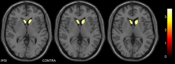
This study included 13 Parkinson's disease patients with peak-of-dose levodopa-induced dyskinesias, 12 nondyskinetic patients, and 12 healthy controls. Participants underwent 5-[I]iodo-3-[2(S)-2-azetidinylmethoxy]pyridine single-photon emission computed tomography, a marker of nicotinic acetylcholine receptors, [I]N--fluoropropyl-2-carbomethoxy-3-(4-iodophenyl)nortropane single-photon emission computed tomography, to measure dopamine reuptake transporter density and 2-[F]fluoro-2-deoxyglucose positron emission tomography to assess regional cerebral metabolic activity. Striatal binding potentials, uptake values at basal ganglia structures, and correlations with clinical variables were analyzed.
Density of nicotinic acetylcholine receptors in the caudate nucleus of dyskinetic subjects was similar to that of healthy controls and significantly higher to that of nondyskinetic patients, in particular, contralaterally to the clinically most affected side.
Our findings support the hypothesis that the expression of dyskinesia may be related to cholinergic neuronal excitability in a dopaminergic-depleted striatum. Cholinergic signaling would play a role in maintaining striatal dopaminergic responsiveness, possibly defining disease phenotype and progression.
研究帕金森病患者左旋多巴诱导的运动障碍与纹状体胆碱能活性之间的关联。
本研究纳入了13例出现剂量峰值左旋多巴诱导运动障碍的帕金森病患者、12例无运动障碍患者以及12名健康对照者。参与者接受了5-[I]碘-3-[2(S)-2-氮杂环丁烷基甲氧基]吡啶单光子发射计算机断层扫描(烟碱型乙酰胆碱受体的标志物)、[I]N-氟丙基-2-甲氧基羰基-3-(4-碘苯基)去甲托烷单光子发射计算机断层扫描以测量多巴胺再摄取转运体密度,以及2-[F]氟-2-脱氧葡萄糖正电子发射断层扫描以评估局部脑代谢活性。分析了纹状体结合潜能、基底节结构的摄取值以及与临床变量的相关性。
运动障碍患者尾状核中烟碱型乙酰胆碱受体的密度与健康对照者相似,且显著高于无运动障碍患者,尤其是在临床受累最严重一侧的对侧。
我们的研究结果支持这样的假说,即运动障碍的表达可能与多巴胺能耗竭的纹状体中的胆碱能神经元兴奋性有关。胆碱能信号传导可能在维持纹状体多巴胺能反应性中发挥作用,这可能决定疾病表型和进展。