• 文献检索
  • 文档翻译
  • 深度研究
  • 学术资讯
  • Suppr Zotero 插件Zotero 插件
  • 邀请有礼
  • 套餐&价格
  • 历史记录
应用&插件
Suppr Zotero 插件Zotero 插件浏览器插件Mac 客户端Windows 客户端微信小程序
定价
高级版会员购买积分包购买API积分包
服务
文献检索文档翻译深度研究API 文档MCP 服务
关于我们
关于 Suppr公司介绍联系我们用户协议隐私条款
关注我们

Suppr 超能文献

核心技术专利:CN118964589B侵权必究
粤ICP备2023148730 号-1Suppr @ 2026

文献检索

告别复杂PubMed语法,用中文像聊天一样搜索,搜遍4000万医学文献。AI智能推荐,让科研检索更轻松。

立即免费搜索

文件翻译

保留排版,准确专业,支持PDF/Word/PPT等文件格式,支持 12+语言互译。

免费翻译文档

深度研究

AI帮你快速写综述,25分钟生成高质量综述,智能提取关键信息,辅助科研写作。

立即免费体验

血肿周围扩散受限是超急性期大脑大血肿的常见表现。

Perihematomal diffusion restriction as a common finding in large intracerebral hemorrhages in the hyperacute phase.

作者信息

Schneider Tanja, Frieling David, Schroeder Julian, Regelsberger Jan, Schoen Gerhard, Fiehler Jens, Gellißen Susanne

机构信息

Department of Diagnostic and Interventional Neuroradiology, University Medical Center Hamburg-Eppendorf, Hamburg, Germany.

Department of Diagnostic and Interventional Radiology, Schön Klinik Hamburg Eilbek, Hamburg, Germany.

出版信息

PLoS One. 2017 Sep 18;12(9):e0184518. doi: 10.1371/journal.pone.0184518. eCollection 2017.

DOI:10.1371/journal.pone.0184518
PMID:28922367
原文链接:https://pmc.ncbi.nlm.nih.gov/articles/PMC5602530/
Abstract

PURPOSE

There is growing evidence that a perihematomal area of restricted diffusion (PDR) exists in intraparenchymal hemorrhages (IPH) within 1 week of symptom onset (SO). Here, we study characteristics and the clinical impact of the PDR in patients with hyperacute (≤ 6 hours from SO) IPH by means of apparent diffusion coefficient (ADC).

METHODS

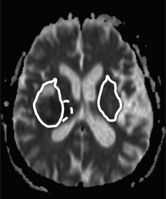
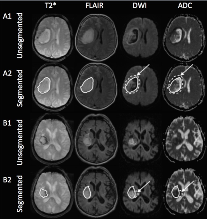
This monocentric, retrospective study includes 83 patients with first-ever primary IPH from 09/2002-10/2015. 3D volumetric segmentation was performed for the IPH, PDR, and perihematomal edema (PHE) on fluid-attenuated inversion recovery, T2*/susceptibility weighted images, and ADC images.

RESULTS

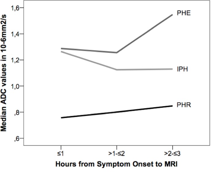
A PDR was seen in 56/83 patients (67.5%) presenting with hyperacute IPH. Multivariate logistic regression analysis revealed every 10-year increase of age (HR 1.929, 95% CI 1.047-3.552, P = .035) and male gender (HR 5.672, 95% CI 1.038-30.992, P = .045) as significant predictors of the presence of a PDR, but not IPH size, IPH location, nor National Institutes of Health Stroke Scale Score (NIHSS) at admission. We found no difference in NIHSS at discharge, hematoma removal, or mortality rate in PDR-positive patients. ADC values of the PDR show a step-wise normalization with increasing time from SO.

CONCLUSIONS

Occurrence of a PDR is a common finding in supratentorial hyperacute IPH, but shows no adverse short-term clinical impact. It may represent transient oligemic and metabolic changes.

摘要

目的

越来越多的证据表明,在症状发作(SO)1周内的脑实质内出血(IPH)中存在血肿周围扩散受限区域(PDR)。在此,我们通过表观扩散系数(ADC)研究超急性(距SO≤6小时)IPH患者中PDR的特征及其临床影响。

方法

这项单中心回顾性研究纳入了2002年9月至2015年10月期间首次发生原发性IPH的83例患者。在液体衰减反转恢复序列、T2* / susceptibility加权图像和ADC图像上对IPH、PDR和血肿周围水肿(PHE)进行三维体积分割。

结果

83例超急性IPH患者中有56例(67.5%)出现PDR。多因素逻辑回归分析显示,年龄每增加10岁(HR 1.929,95%CI 1.047 - 3.552,P = 0.035)和男性(HR 5.672,95%CI 1.038 - 30.992,P = 0.045)是PDR存在的显著预测因素,但不是IPH大小、IPH位置或入院时的美国国立卫生研究院卒中量表评分(NIHSS)。我们发现PDR阳性患者出院时的NIHSS、血肿清除情况或死亡率没有差异。PDR的ADC值随距SO时间增加呈逐步正常化。

结论

PDR的出现是幕上超急性IPH中的常见表现,但在短期临床中没有不良影响。它可能代表短暂的低灌注和代谢变化。

https://cdn.ncbi.nlm.nih.gov/pmc/blobs/b394/5602530/390ca67fa701/pone.0184518.g003.jpg
https://cdn.ncbi.nlm.nih.gov/pmc/blobs/b394/5602530/5ad575dedcab/pone.0184518.g001.jpg
https://cdn.ncbi.nlm.nih.gov/pmc/blobs/b394/5602530/ae852f18f7a9/pone.0184518.g002.jpg
https://cdn.ncbi.nlm.nih.gov/pmc/blobs/b394/5602530/390ca67fa701/pone.0184518.g003.jpg
https://cdn.ncbi.nlm.nih.gov/pmc/blobs/b394/5602530/5ad575dedcab/pone.0184518.g001.jpg
https://cdn.ncbi.nlm.nih.gov/pmc/blobs/b394/5602530/ae852f18f7a9/pone.0184518.g002.jpg
https://cdn.ncbi.nlm.nih.gov/pmc/blobs/b394/5602530/390ca67fa701/pone.0184518.g003.jpg

相似文献

1
Perihematomal diffusion restriction as a common finding in large intracerebral hemorrhages in the hyperacute phase.血肿周围扩散受限是超急性期大脑大血肿的常见表现。
PLoS One. 2017 Sep 18;12(9):e0184518. doi: 10.1371/journal.pone.0184518. eCollection 2017.
2
Perihematomal Diffusion Restriction in Intracerebral Hemorrhage Depends on Hematoma Volume, But Does Not Predict Outcome.脑出血周围的扩散受限取决于血肿体积,但不能预测预后。
Cerebrovasc Dis. 2016;42(3-4):280-7. doi: 10.1159/000446549. Epub 2016 May 25.
3
Diffusion-perfusion MR evaluation of perihematomal injury in hyperacute intracerebral hemorrhage.超急性脑出血血肿周围损伤的扩散灌注磁共振成像评估
Neurology. 2001 Nov 13;57(9):1611-7. doi: 10.1212/wnl.57.9.1611.
4
Perihematomal edema in primary intracerebral hemorrhage is plasma derived.原发性脑出血中的血肿周围水肿源自血浆。
Stroke. 2004 Aug;35(8):1879-85. doi: 10.1161/01.STR.0000131807.54742.1a. Epub 2004 Jun 3.
5
Rate of Perihematomal Edema Expansion Predicts Outcome After Intracerebral Hemorrhage.血肿周围水肿扩展速率可预测脑出血后的结局。
Crit Care Med. 2016 Apr;44(4):790-7. doi: 10.1097/CCM.0000000000001553.
6
Temporal pattern of cytotoxic edema in the perihematomal region after intracerebral hemorrhage: a serial magnetic resonance imaging study.脑出血后血肿周围区细胞毒性水肿的时间模式:一项连续磁共振成像研究。
Stroke. 2013 Apr;44(4):1144-6. doi: 10.1161/STROKEAHA.111.000056. Epub 2013 Feb 7.
7
Natural history of perihematomal edema after intracerebral hemorrhage measured by serial magnetic resonance imaging.脑出血后磁共振成像序列测量的血肿周围水肿自然史。
Stroke. 2011 Jan;42(1):73-80. doi: 10.1161/STROKEAHA.110.590646. Epub 2010 Dec 16.
8
Prognostic significance of perihematomal edema in acute intracerebral hemorrhage: pooled analysis from the intensive blood pressure reduction in acute cerebral hemorrhage trial studies.血肿周围水肿对急性脑出血的预后意义:强化降压治疗急性脑出血试验研究的荟萃分析。
Stroke. 2015 Apr;46(4):1009-13. doi: 10.1161/STROKEAHA.114.007154. Epub 2015 Feb 24.
9
Diffusion-weighted MR imaging of intracerebral hemorrhage.脑内出血的扩散加权磁共振成像
Korean J Radiol. 2001 Oct-Dec;2(4):183-91. doi: 10.3348/kjr.2001.2.4.183.
10
Perihematomal Edema Is Greater in the Presence of a Spot Sign but Does Not Predict Intracerebral Hematoma Expansion.血肿周围水肿在存在斑点征时更明显,但不能预测颅内血肿扩大。
Stroke. 2016 Feb;47(2):350-5. doi: 10.1161/STROKEAHA.115.011295. Epub 2015 Dec 22.

引用本文的文献

1
Molecular, Pathological, Clinical, and Therapeutic Aspects of Perihematomal Edema in Different Stages of Intracerebral Hemorrhage.血肿不同阶段的血肿周围水肿的分子、病理、临床和治疗方面。
Oxid Med Cell Longev. 2022 Sep 17;2022:3948921. doi: 10.1155/2022/3948921. eCollection 2022.

本文引用的文献

1
Perihematomal Diffusion Restriction in Intracerebral Hemorrhage Depends on Hematoma Volume, But Does Not Predict Outcome.脑出血周围的扩散受限取决于血肿体积,但不能预测预后。
Cerebrovasc Dis. 2016;42(3-4):280-7. doi: 10.1159/000446549. Epub 2016 May 25.
2
Diffusion-weighted signal patterns of intracranial haemorrhage.颅内出血的扩散加权信号模式。
Clin Radiol. 2015 Aug;70(8):909-16. doi: 10.1016/j.crad.2015.04.006. Epub 2015 Jun 4.
3
Quality assurance multicenter comparison of different MR scanners for quantitative diffusion-weighted imaging.
不同磁共振成像(MR)扫描仪用于定量扩散加权成像的质量保证多中心比较
J Magn Reson Imaging. 2016 Jan;43(1):213-9. doi: 10.1002/jmri.24956. Epub 2015 May 26.
4
Ischemia in intracerebral hemorrhage is associated with leukoaraiosis and hematoma volume, not blood pressure reduction.脑出血中的缺血与脑白质疏松症和血肿体积相关,而非与血压降低相关。
Stroke. 2015 Jun;46(6):1541-7. doi: 10.1161/STROKEAHA.114.008304. Epub 2015 Apr 28.
5
Temporal pattern of cytotoxic edema in the perihematomal region after intracerebral hemorrhage: a serial magnetic resonance imaging study.脑出血后血肿周围区细胞毒性水肿的时间模式:一项连续磁共振成像研究。
Stroke. 2013 Apr;44(4):1144-6. doi: 10.1161/STROKEAHA.111.000056. Epub 2013 Feb 7.
6
Temporal changes in perihematomal apparent diffusion coefficient values during the transition from acute to subacute phases in patients with spontaneous intracerebral hemorrhage.自发性脑出血患者从急性期向亚急性期过渡期间血肿周围表观弥散系数值的时间变化。
Neuroradiology. 2013 Feb;55(2):145-56. doi: 10.1007/s00234-012-1093-x. Epub 2012 Sep 18.
7
MRI profile of the perihematomal region in acute intracerebral hemorrhage.急性脑出血血肿周围区域的 MRI 特征。
Stroke. 2010 Nov;41(11):2681-3. doi: 10.1161/STROKEAHA.110.590638. Epub 2010 Oct 14.
8
Development and validation of a simple conversion model for comparison of intracerebral hemorrhage volumes measured on CT and gradient recalled echo MRI.一种用于比较CT和梯度回波MRI测量的脑出血体积的简单转换模型的开发与验证
Stroke. 2008 Jul;39(7):2017-20. doi: 10.1161/STROKEAHA.107.505719. Epub 2008 May 15.
9
Evolution of early perihemorrhagic changes--ischemia vs. edema: an MRI study in rats.早期出血周围改变的演变——缺血与水肿:大鼠的MRI研究
Exp Neurol. 2005 Jun;193(2):369-76. doi: 10.1016/j.expneurol.2005.01.017.
10
Spontaneous intracerebral hematoma on diffusion-weighted images: influence of T2-shine-through and T2-blackout effects.扩散加权成像上的自发性脑内血肿:T2透过效应和T2消影效应的影响
AJNR Am J Neuroradiol. 2005 Feb;26(2):236-41.