Buxboim Amnon, Irianto Jerome, Swift Joe, Athirasala Avathamsa, Shin Jae-Won, Rehfeldt Florian, Discher Dennis E
Molecular and Cell Biophysics Laboratory, University of Pennsylvania, Philadelphia, PA 19104.
Department/Graduate Group of Physics and Astronomy, University of Pennsylvania, Philadelphia, PA 19104.
Mol Biol Cell. 2017 Nov 7;28(23):3333-3348. doi: 10.1091/mbc.E17-06-0393. Epub 2017 Sep 20.
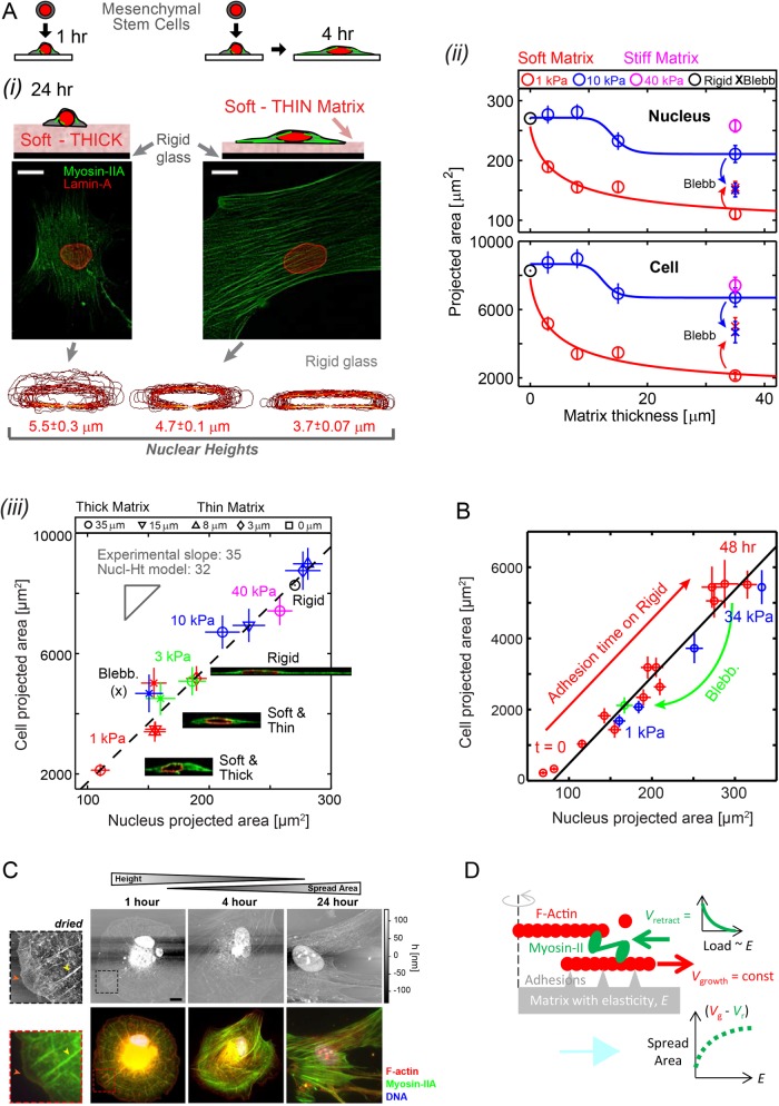
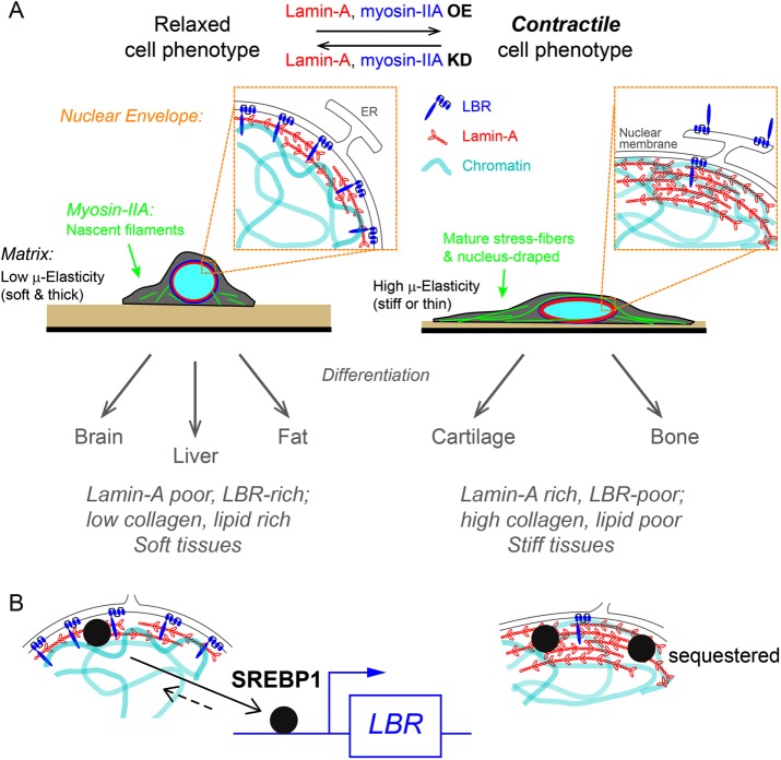
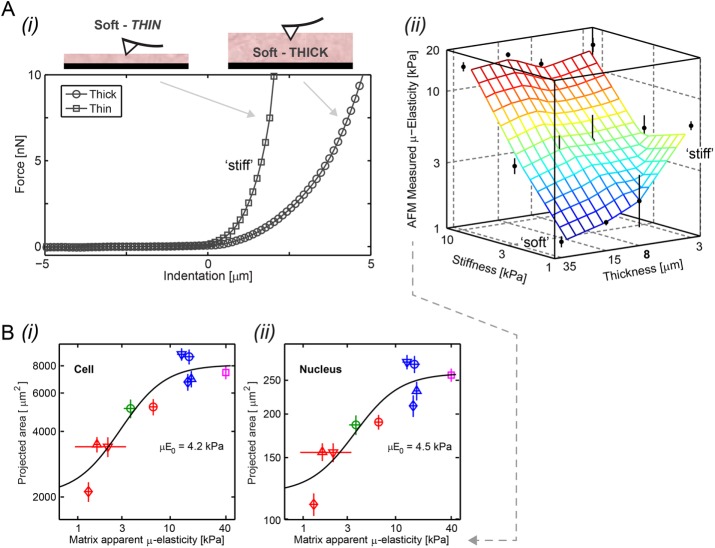
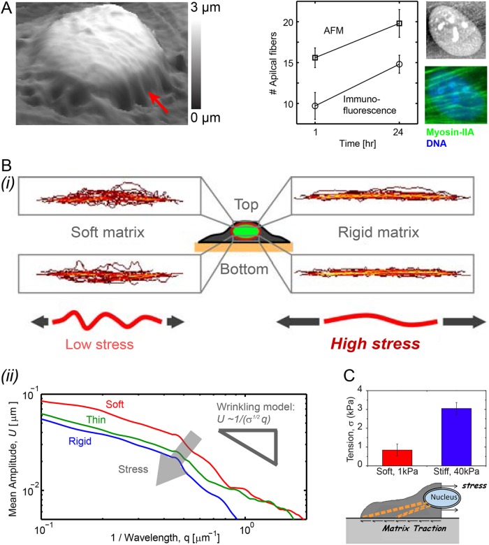
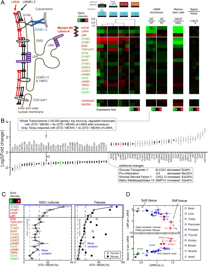
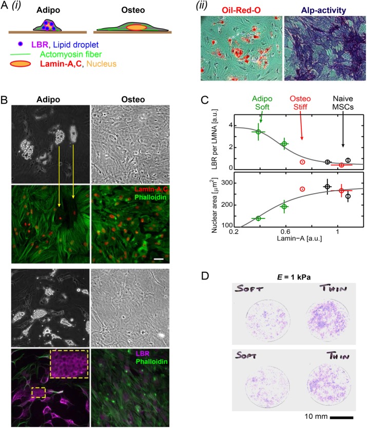
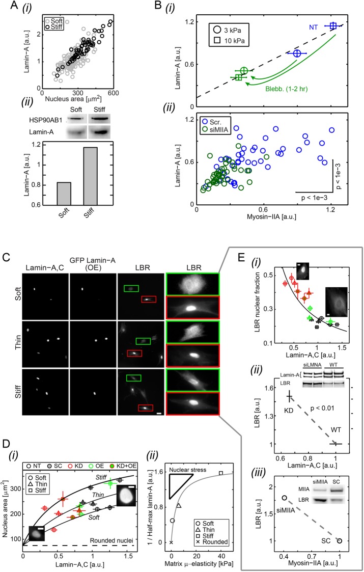
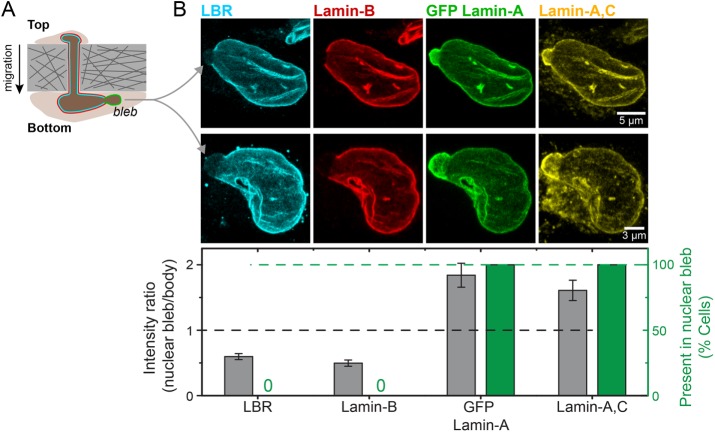
Matrix stiffness that is sensed by a cell or measured by a purely physical probe reflects the intrinsic elasticity of the matrix and also how thick or thin the matrix is. Here, mesenchymal stem cells (MSCs) and their nuclei spread in response to thickness-corrected matrix microelasticity, with increases in nuclear tension and nuclear stiffness resulting from increases in myosin-II and lamin-A,C. Linearity between the widely varying projected area of a cell and its nucleus across many matrices, timescales, and myosin-II activity levels indicates a constant ratio of nucleus-to-cell volume, despite MSCs' lineage plasticity. Nuclear envelope fluctuations are suppressed on the stiffest matrices, and fluctuation spectra reveal a high nuclear tension that matches trends from traction force microscopy and from increased lamin-A,C. Transcriptomes of many diverse tissues and MSCs further show that lamin-A,C's increase with tissue or matrix stiffness anti-correlates with lamin-B receptor (LBR), which contributes to lipid/sterol biosynthesis. Adipogenesis (a soft lineage) indeed increases LBR:lamin-A,C protein stoichiometry in MSCs versus osteogenesis (stiff). The two factors compete for lamin-B in response to matrix elasticity, knockdown, myosin-II inhibition, and even constricted migration that disrupts and segregates lamins in situ. Matrix stiffness-driven contractility thus tenses the nucleus to favor lamin-A,C accumulation and suppress soft tissue phenotypes.
细胞感知到的或通过纯物理探针测量的基质硬度反映了基质的固有弹性以及基质的厚度。在这里,间充质干细胞(MSCs)及其细胞核会随着经厚度校正的基质微弹性而展开,肌球蛋白-II和核纤层蛋白A、C的增加导致核张力和核硬度增加。在许多基质、时间尺度和肌球蛋白-II活性水平上,细胞及其细胞核的广泛变化的投影面积之间的线性关系表明,尽管间充质干细胞具有谱系可塑性,但细胞核与细胞体积的比例是恒定的。在最硬的基质上,核膜波动受到抑制,波动光谱显示出高核张力,这与牵引力显微镜以及核纤层蛋白A、C增加的趋势相匹配。许多不同组织和间充质干细胞的转录组进一步表明,核纤层蛋白A、C随组织或基质硬度的增加与参与脂质/固醇生物合成的核纤层蛋白B受体(LBR)呈负相关。事实上,与成骨(硬谱系)相比,脂肪生成(软谱系)会增加间充质干细胞中LBR与核纤层蛋白A、C的蛋白质化学计量比。这两个因素在响应基质弹性、基因敲除、肌球蛋白-II抑制甚至干扰和原位分离核纤层蛋白的受限迁移时,会竞争核纤层蛋白B。因此,基质硬度驱动的收缩性使细胞核紧张,有利于核纤层蛋白A、C的积累并抑制软组织表型。