Aab Cardiovascular Research Institute, Department of Medicine, University of Rochester School of Medicine and Dentistry, Rochester, NY, USA.
Department of Biomedical Genetics, University of Rochester School of Medicine and Dentistry, Rochester, NY, USA.
Sci Rep. 2017 Sep 21;7(1):12081. doi: 10.1038/s41598-017-12321-7.
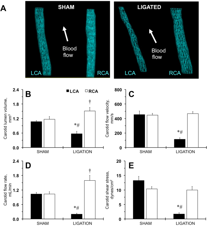
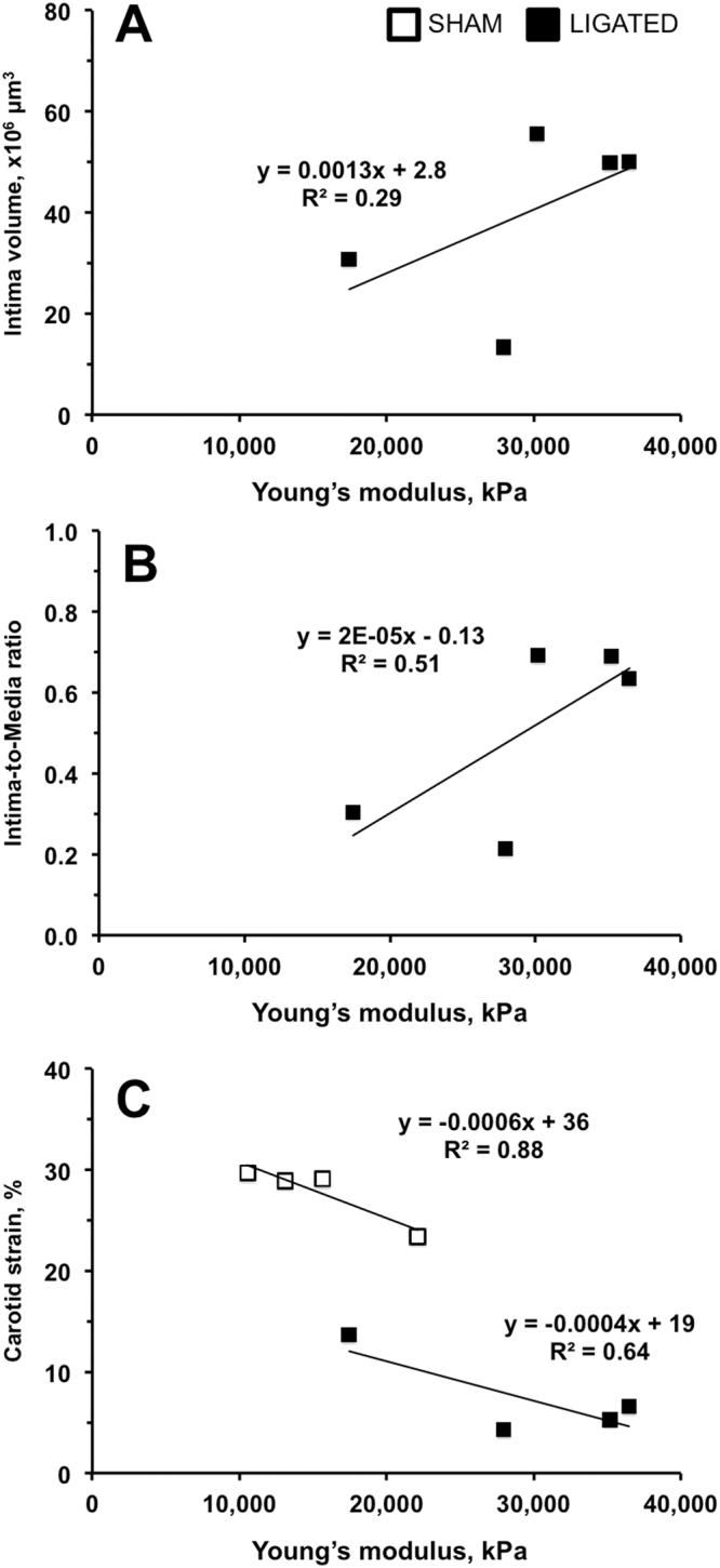
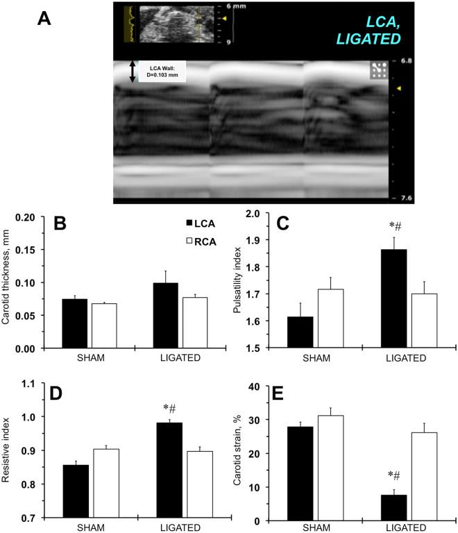
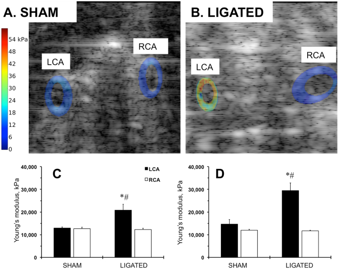
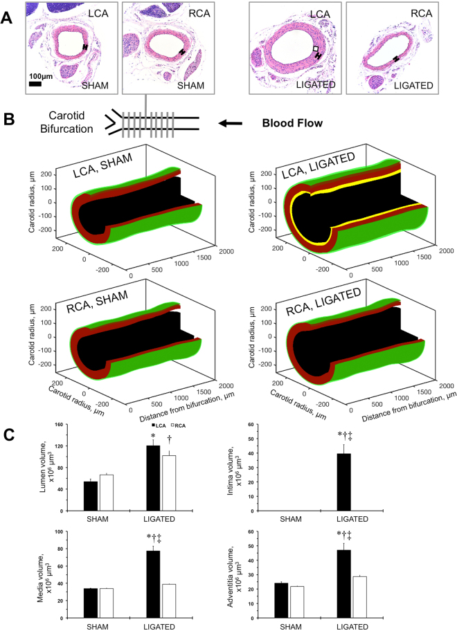
Increased arterial thickness measured with ultrasound correlates with future cardiovascular events, but conventional ultrasound imaging techniques cannot distinguish between intima, media, or atherosclerotic plaque in the carotid artery. In this work, we evaluated how well vascular elastography can detect intimal changes in a mouse model of carotid remodeling. We ligated the left external and internal branches of the carotid artery of male FVB mice and performed sham operations for 2 weeks. High-resolution ultrasound imaging accurately detected lower blood velocities and low blood volume flow in the carotid arteries after ligation in FVB mice. However, ultrasound could not detect differences in the carotid wall even at 2 weeks post-surgery. The Young's modulus was measured based on displacements of the carotid artery wall, and Young's modulus was 2-fold greater in shams at 1 week post ligation, and 3-fold greater 2 weeks after ligation. Finally, the higher Young's modulus was most associated with higher intimal thickness but not medial or adventitial thickness as measured by histology. In conclusion, we developed a robust ultrasound-based elastography method for early detection of intimal changes in small animals.
用超声测量的动脉壁增厚与未来的心血管事件相关,但传统的超声成像技术无法区分颈动脉的内膜、中膜或粥样硬化斑块。在这项工作中,我们评估了血管弹性成像技术在颈动脉重塑的小鼠模型中检测内膜变化的能力。我们结扎雄性 FVB 小鼠的颈外动脉和颈内动脉的分支,并进行 2 周的假手术。高分辨率超声成像准确地检测到 FVB 小鼠结扎后颈动脉内血流速度降低和血流量减少。然而,即使在手术后 2 周,超声也无法检测到颈动脉壁的差异。根据颈动脉壁的位移测量杨氏模量,结扎后 1 周,假手术组的杨氏模量增加了 2 倍,结扎后 2 周增加了 3 倍。最后,较高的杨氏模量与内膜厚度的增加最相关,但与组织学测量的中膜或外膜厚度无关。总之,我们开发了一种基于超声的弹性成像方法,用于早期检测小动物的内膜变化。