von Furstenberg Richard J, Li Joy, Stolarchuk Christina, Feder Rachel, Campbell Alexa, Kruger Leandi, Gonzalez Liara M, Blikslager Anthony T, Cardona Diana M, McCall Shannon J, Henning Susan J, Garman Katherine S
Division of Gastroenterology, Department of Medicine, Duke University, Durham, North Carolina.
Department of Clinical Sciences, North Carolina State University, College of Veterinary Medicine, Raleigh, North Carolina.
Cell Mol Gastroenterol Hepatol. 2017 Aug 4;4(3):385-404. doi: 10.1016/j.jcmgh.2017.07.005. eCollection 2017 Nov.
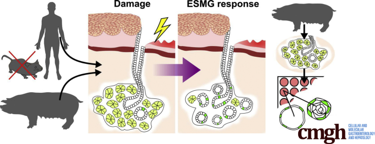
BACKGROUND & AIMS: Although cells comprising esophageal submucosal glands (ESMGs) represent a potential progenitor cell niche, new models are needed to understand their capacity to proliferate and differentiate. By histologic appearance, ESMGs have been associated with both overlying normal squamous epithelium and columnar epithelium. Our aim was to assess ESMG proliferation and differentiation in a 3-dimensional culture model.
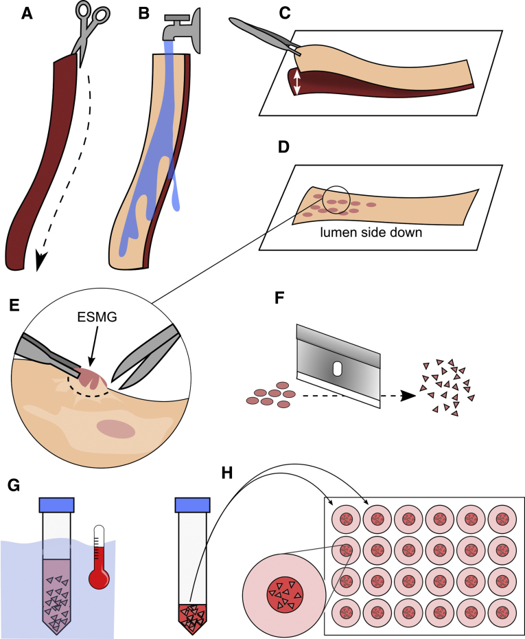
We evaluated proliferation in human ESMGs from normal and diseased tissue by proliferating cell nuclear antigen immunohistochemistry. Next, we compared 5-ethynyl-2'-deoxyuridine labeling in porcine ESMGs in vivo before and after esophageal injury with a novel in vitro porcine organoid ESMG model. Microarray analysis of ESMGs in culture was compared with squamous epithelium and fresh ESMGs.
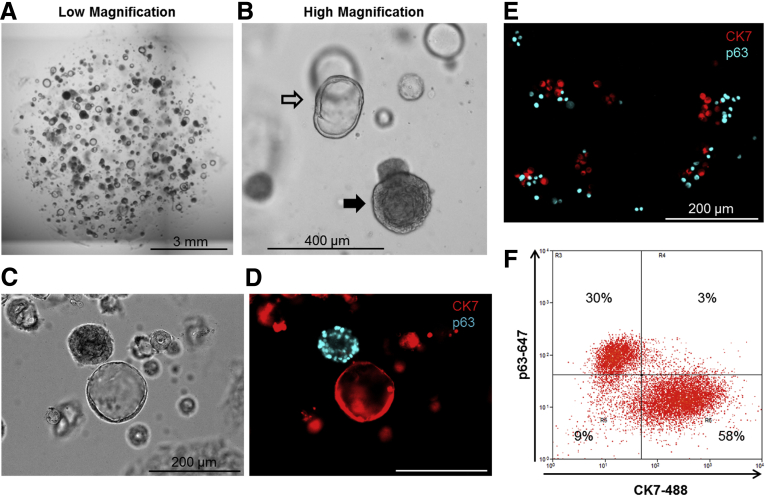
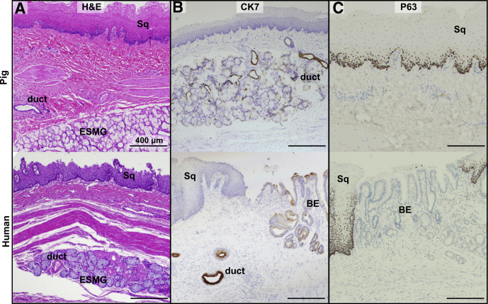
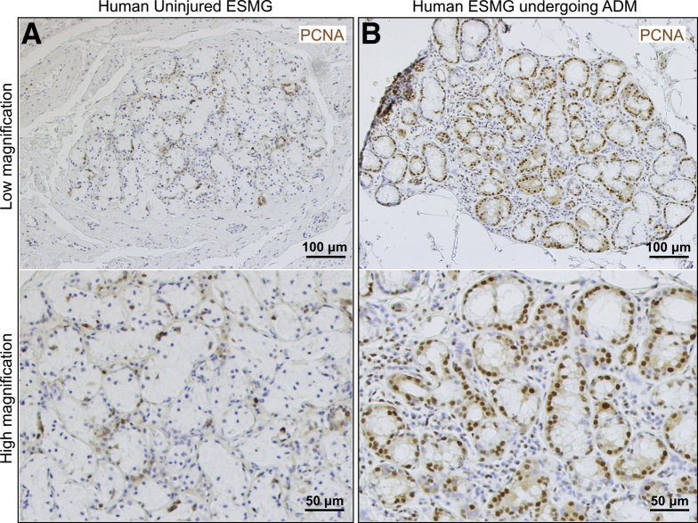
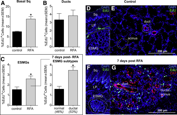
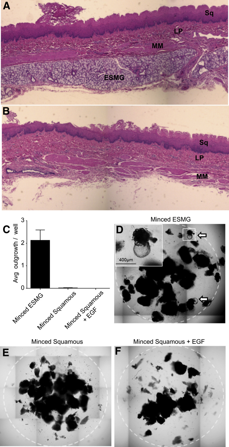
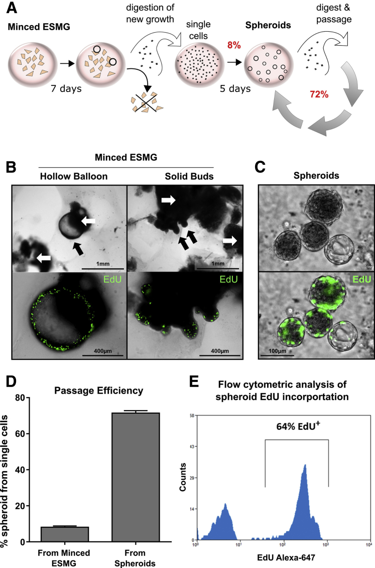
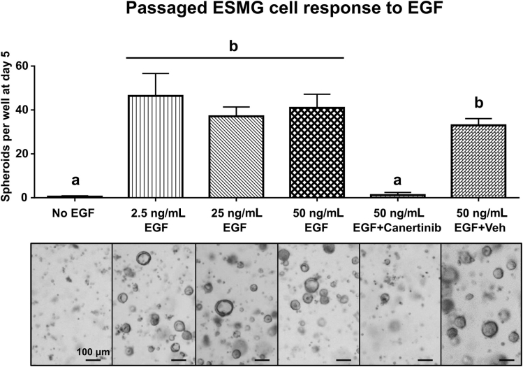
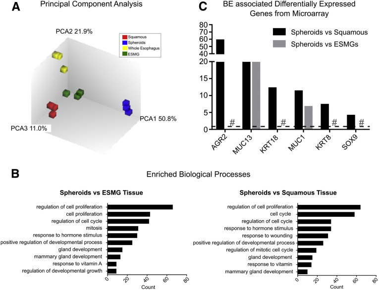
Marked proliferation was observed in human ESMGs of diseased tissue. This activated ESMG state was recapitulated after esophageal injury in an in vivo porcine model, ESMGs assumed a ductal appearance with increased proliferation compared with control. Isolated and cultured porcine ESMGs produced buds with actively cycling cells and passaged to form epidermal growth factor-dependent spheroids. These spheroids were highly proliferative and were passaged multiple times. Two phenotypes of spheroids were identified: solid squamous (P63+) and hollow/ductal (cytokeratin 7+). Microarray analysis showed spheroids to be distinct from parent ESMGs and enriched for columnar transcripts.
Our results suggest that the activated ESMG state, seen in both human disease and our porcine model, may provide a source of cells to repopulate damaged epithelium in a normal manner (squamous) or abnormally (columnar epithelium). This culture model will allow the evaluation of factors that drive ESMGs in the regeneration of injured epithelium. The raw microarray data have been uploaded to the National Center for Biotechnology Information Gene Expression Omnibus (accession number: GSE100543).
尽管构成食管黏膜下腺(ESMGs)的细胞代表了一个潜在的祖细胞龛,但仍需要新的模型来了解它们的增殖和分化能力。从组织学外观来看,ESMGs与覆盖其上的正常鳞状上皮和柱状上皮均有关联。我们的目的是在三维培养模型中评估ESMGs的增殖和分化。
我们通过增殖细胞核抗原免疫组织化学评估来自正常和病变组织的人ESMGs中的增殖情况。接下来,我们将猪食管损伤前后体内ESMGs中的5-乙炔基-2'-脱氧尿苷标记与一种新型的体外猪类器官ESMG模型进行比较。将培养的ESMGs的微阵列分析结果与鳞状上皮和新鲜ESMGs进行比较。
在病变组织的人ESMGs中观察到明显的增殖。在体内猪模型中,食管损伤后再现了这种活化的ESMG状态,与对照组相比,ESMGs呈现导管样外观且增殖增加。分离并培养的猪ESMGs产生了带有活跃循环细胞的芽,并传代形成表皮生长因子依赖性球体。这些球体具有高度增殖性,并能传代多次。鉴定出两种球体表型:实性鳞状(P63+)和中空/导管样(细胞角蛋白7+)。微阵列分析表明,球体与亲本ESMGs不同,且富含柱状转录本。
我们 的结果表明,在人类疾病和我们的猪模型中均可见的活化ESMG状态,可能提供细胞来源以正常方式(鳞状)或异常方式(柱状上皮)重新填充受损上皮。这种培养模型将有助于评估驱动ESMGs参与损伤上皮再生的因素。原始微阵列数据已上传至美国国立生物技术信息中心基因表达综合数据库(登录号:GSE100543)。