• 文献检索
  • 文档翻译
  • 深度研究
  • 学术资讯
  • Suppr Zotero 插件Zotero 插件
  • 邀请有礼
  • 套餐&价格
  • 历史记录
应用&插件
Suppr Zotero 插件Zotero 插件浏览器插件Mac 客户端Windows 客户端微信小程序
定价
高级版会员购买积分包购买API积分包
服务
文献检索文档翻译深度研究API 文档MCP 服务
关于我们
关于 Suppr公司介绍联系我们用户协议隐私条款
关注我们

Suppr 超能文献

核心技术专利:CN118964589B侵权必究
粤ICP备2023148730 号-1Suppr @ 2026

文献检索

告别复杂PubMed语法,用中文像聊天一样搜索,搜遍4000万医学文献。AI智能推荐,让科研检索更轻松。

立即免费搜索

文件翻译

保留排版,准确专业,支持PDF/Word/PPT等文件格式,支持 12+语言互译。

免费翻译文档

深度研究

AI帮你快速写综述,25分钟生成高质量综述,智能提取关键信息,辅助科研写作。

立即免费体验

双面显微镜实现药用蛭整体神经节膜电位成像。

A double-sided microscope to realize whole-ganglion imaging of membrane potential in the medicinal leech.

机构信息

Division of Biology and Biological Engineering, California Institute of Technology, Pasadena, United States.

出版信息

Elife. 2017 Sep 25;6:e29839. doi: 10.7554/eLife.29839.

DOI:10.7554/eLife.29839
PMID:28944754
原文链接:https://pmc.ncbi.nlm.nih.gov/articles/PMC5656430/
Abstract

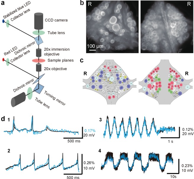
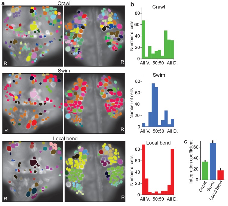
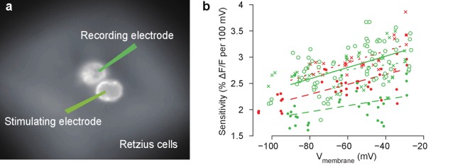
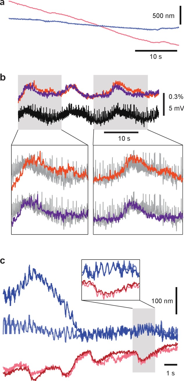
Studies of neuronal network emergence during sensory processing and motor control are greatly facilitated by technologies that allow us to simultaneously record the membrane potential dynamics of a large population of neurons in single cell resolution. To achieve whole-brain recording with the ability to detect both small synaptic potentials and action potentials, we developed a voltage-sensitive dye (VSD) imaging technique based on a double-sided microscope that can image two sides of a nervous system simultaneously. We applied this system to the segmental ganglia of the medicinal leech. Double-sided VSD imaging enabled simultaneous recording of membrane potential events from almost all of the identifiable neurons. Using data obtained from double-sided VSD imaging, we analyzed neuronal dynamics in both sensory processing and generation of behavior and constructed functional maps for identification of neurons contributing to these processes.

摘要

在感官处理和运动控制过程中,神经元网络的出现研究极大地受益于能够以单细胞分辨率同时记录大量神经元膜电位动力学的技术。为了实现具有检测小突触电位和动作电位能力的全脑记录,我们开发了一种基于双面显微镜的电压敏感染料 (VSD) 成像技术,该技术能够同时对神经系统的两面进行成像。我们将该系统应用于医用水蛭的节段性神经节。双面 VSD 成像使我们能够同时记录几乎所有可识别神经元的膜电位事件。利用从双面 VSD 成像获得的数据,我们分析了感觉处理和行为产生过程中的神经元动力学,并构建了功能图谱以鉴定对这些过程有贡献的神经元。

https://cdn.ncbi.nlm.nih.gov/pmc/blobs/4ef0/5656430/142c39ffd0eb/elife-29839-fig5.jpg
https://cdn.ncbi.nlm.nih.gov/pmc/blobs/4ef0/5656430/d3b073f7b80c/elife-29839-fig1.jpg
https://cdn.ncbi.nlm.nih.gov/pmc/blobs/4ef0/5656430/1d76383eb968/elife-29839-fig1-figsupp1.jpg
https://cdn.ncbi.nlm.nih.gov/pmc/blobs/4ef0/5656430/feaa6d329f3b/elife-29839-fig1-figsupp2.jpg
https://cdn.ncbi.nlm.nih.gov/pmc/blobs/4ef0/5656430/123903a55b0f/elife-29839-fig1-figsupp3.jpg
https://cdn.ncbi.nlm.nih.gov/pmc/blobs/4ef0/5656430/eb61b596f7aa/elife-29839-fig2.jpg
https://cdn.ncbi.nlm.nih.gov/pmc/blobs/4ef0/5656430/8f5433379a46/elife-29839-fig2-figsupp1.jpg
https://cdn.ncbi.nlm.nih.gov/pmc/blobs/4ef0/5656430/3724840a6284/elife-29839-fig3.jpg
https://cdn.ncbi.nlm.nih.gov/pmc/blobs/4ef0/5656430/1f20a6101dae/elife-29839-fig3-figsupp1.jpg
https://cdn.ncbi.nlm.nih.gov/pmc/blobs/4ef0/5656430/92819d227def/elife-29839-fig4.jpg
https://cdn.ncbi.nlm.nih.gov/pmc/blobs/4ef0/5656430/142c39ffd0eb/elife-29839-fig5.jpg

相似文献

1
A double-sided microscope to realize whole-ganglion imaging of membrane potential in the medicinal leech.双面显微镜实现药用蛭整体神经节膜电位成像。
Elife. 2017 Sep 25;6:e29839. doi: 10.7554/eLife.29839.
2
Simultaneous measurement of membrane potential changes in multiple pattern generating neurons using voltage sensitive dye imaging.使用电压敏感染料成像技术同时测量多个模式生成神经元的膜电位变化。
J Neurosci Methods. 2012 Jan 15;203(1):78-88. doi: 10.1016/j.jneumeth.2011.09.015. Epub 2011 Sep 22.
3
Estimation of neuronal activity based on voltage-sensitive dye imaging in a moving preparation.基于电压敏感染料成像在运动标本中估计神经元活动。
Annu Int Conf IEEE Eng Med Biol Soc. 2015;2015:6285-8. doi: 10.1109/EMBC.2015.7319829.
4
A classic model animal in the 21st century: recent lessons from the leech nervous system.21世纪的经典模式动物:来自水蛭神经系统的最新经验教训。
J Exp Biol. 2015 Nov;218(Pt 21):3353-9. doi: 10.1242/jeb.113860.
5
Single-sweep voltage-sensitive dye imaging of interacting identified neurons.单扫描电压敏感染料成像技术在相互作用的鉴定神经元中的应用。
J Neurosci Methods. 2011 Jan 15;194(2):224-34. doi: 10.1016/j.jneumeth.2010.10.007. Epub 2010 Oct 20.
6
Monitoring Spiking Activity of Many Individual Neurons in Invertebrate Ganglia.监测无脊椎动物神经节中多个单个神经元的放电活动。
Adv Exp Med Biol. 2015;859:127-45. doi: 10.1007/978-3-319-17641-3_5.
7
Dual-sided Voltage-sensitive Dye Imaging of Leech Ganglia.水蛭神经节的双侧电压敏感染料成像
Bio Protoc. 2018 Mar 5;8(5). doi: 10.21769/BioProtoc.2751.
8
Monitoring Integrated Activity of Individual Neurons Using FRET-Based Voltage-Sensitive Dyes.使用基于荧光共振能量转移(FRET)的电压敏感染料监测单个神经元的综合活动。
Adv Exp Med Biol. 2015;859:149-69. doi: 10.1007/978-3-319-17641-3_6.
9
Glial responses during evoked behaviors in the leech.水蛭诱发行为期间的神经胶质反应。
Glia. 1999 Apr;26(2):186-9. doi: 10.1002/(sici)1098-1136(199904)26:2<186::aid-glia9>3.0.co;2-e.
10
Temporal correlation between neuronal tail ganglion activity and locomotion in the leech, Hirudo medicinalis.药用水蛭(Hirudo medicinalis)中神经元尾神经节活动与运动之间的时间相关性。
Invert Neurosci. 1997 Mar;2(4):245-51. doi: 10.1007/BF02211937.

引用本文的文献

1
Cell anatomy and network input explain differences within but not between leech touch cells at two different locations.细胞解剖结构和网络输入解释了同一水蛭不同位置触觉细胞之间而非不同水蛭触觉细胞之间的差异。
Front Cell Neurosci. 2023 Jul 25;17:1186997. doi: 10.3389/fncel.2023.1186997. eCollection 2023.
2
Current Practice in Using Voltage Imaging to Record Fast Neuronal Activity: Successful Examples from Invertebrate to Mammalian Studies.当前使用电压成像记录快速神经元活动的实践:无脊椎动物到哺乳动物研究中的成功案例。
Biosensors (Basel). 2023 Jun 13;13(6):648. doi: 10.3390/bios13060648.
3
Initial Variability and Time-Dependent Changes of Neuronal Response Features Are Cell-Type-Specific.

本文引用的文献

1
Dual-sided Voltage-sensitive Dye Imaging of Leech Ganglia.水蛭神经节的双侧电压敏感染料成像
Bio Protoc. 2018 Mar 5;8(5). doi: 10.21769/BioProtoc.2751.
2
Neural Circuits Underlying Fly Larval Locomotion.果蝇幼虫运动的神经回路
Curr Pharm Des. 2017;23(12):1722-1733. doi: 10.2174/1381612822666161208120835.
3
Encoding of Tactile Stimuli by Mechanoreceptors and Interneurons of the Medicinal Leech.药用水蛭的机械感受器和中间神经元对触觉刺激的编码
神经元反应特征的初始变异性和时间依赖性变化具有细胞类型特异性。
Front Cell Neurosci. 2022 Apr 27;16:858221. doi: 10.3389/fncel.2022.858221. eCollection 2022.
4
Voltage imaging identifies spinal circuits that modulate locomotor adaptation in zebrafish.电压成像可识别调节斑马鱼运动适应的脊髓回路。
Neuron. 2022 Apr 6;110(7):1211-1222.e4. doi: 10.1016/j.neuron.2022.01.001. Epub 2022 Jan 31.
5
Anatomy and activity patterns in a multifunctional motor neuron and its surrounding circuits.多功能运动神经元及其周围回路的解剖结构和活动模式。
Elife. 2021 Feb 15;10:e61881. doi: 10.7554/eLife.61881.
6
Photodiode-Based Optical Imaging for Recording Network Dynamics with Single-Neuron Resolution in Non-Transgenic Invertebrates.基于光电二极管的光学成像技术,用于在非转基因无脊椎动物中以单神经元分辨率记录网络动态。
J Vis Exp. 2020 Jul 9(161). doi: 10.3791/61623.
7
Monitoring neuronal activity with voltage-sensitive fluorophores.使用电压敏感染料监测神经元活动。
Methods Enzymol. 2020;640:185-204. doi: 10.1016/bs.mie.2020.04.028. Epub 2020 Jun 1.
8
Brief Electrical Stimulation Triggers an Effective Regeneration of Leech CNS.短暂电刺激可有效触发医用水蛭 CNS 再生。
eNeuro. 2020 Jun 25;7(3). doi: 10.1523/ENEURO.0030-19.2020. Print 2020 May/Jun.
9
Electrophysiology, Unplugged: Imaging Membrane Potential with Fluorescent Indicators.电生理学, unplugged:用荧光指示剂成像膜电位。
Acc Chem Res. 2020 Jan 21;53(1):11-19. doi: 10.1021/acs.accounts.9b00514. Epub 2019 Dec 13.
10
Dual-sided Voltage-sensitive Dye Imaging of Leech Ganglia.水蛭神经节的双侧电压敏感染料成像
Bio Protoc. 2018 Mar 5;8(5). doi: 10.21769/BioProtoc.2751.
Front Physiol. 2016 Oct 28;7:506. doi: 10.3389/fphys.2016.00506. eCollection 2016.
4
Patterns and distribution of presynaptic and postsynaptic elements within serial electron microscopic reconstructions of neuronal arbors from the medicinal leech Hirudo verbana.来自药用蛭(Hirudo verbana)神经元树突连续电子显微镜重建中突触前和突触后元件的模式与分布。
J Comp Neurol. 2016 Dec 15;524(18):3677-3695. doi: 10.1002/cne.24120.
5
Scalable Semisupervised Functional Neurocartography Reveals Canonical Neurons in Behavioral Networks.可扩展的半监督功能神经图谱揭示行为网络中的典型神经元。
Neural Comput. 2016 Aug;28(8):1453-97. doi: 10.1162/NECO_a_00852. Epub 2016 Jun 27.
6
Whole-brain calcium imaging with cellular resolution in freely behaving Caenorhabditis elegans.在自由活动的秀丽隐杆线虫中进行具有细胞分辨率的全脑钙成像。
Proc Natl Acad Sci U S A. 2016 Feb 23;113(8):E1074-81. doi: 10.1073/pnas.1507110112. Epub 2015 Dec 28.
7
Memory Formation in Tritonia via Recruitment of Variably Committed Neurons.通过募集不同程度定向分化的神经元在三角涡虫中形成记忆
Curr Biol. 2015 Nov 16;25(22):2879-88. doi: 10.1016/j.cub.2015.09.033. Epub 2015 Nov 5.
8
Global brain dynamics embed the motor command sequence of Caenorhabditis elegans.全球大脑动力学嵌入秀丽隐杆线虫的运动指令序列。
Cell. 2015 Oct 22;163(3):656-69. doi: 10.1016/j.cell.2015.09.034. Epub 2015 Oct 17.
9
Modular deconstruction reveals the dynamical and physical building blocks of a locomotion motor program.模块化解构揭示了运动运动程序的动态和物理构建块。
Neuron. 2015 Apr 8;86(1):304-18. doi: 10.1016/j.neuron.2015.03.005. Epub 2015 Mar 26.
10
Responses to conflicting stimuli in a simple stimulus-response pathway.在简单刺激-反应通路中对冲突刺激的反应。
J Neurosci. 2015 Feb 11;35(6):2398-406. doi: 10.1523/JNEUROSCI.3823-14.2015.