• 文献检索
  • 文档翻译
  • 深度研究
  • 学术资讯
  • Suppr Zotero 插件Zotero 插件
  • 邀请有礼
  • 套餐&价格
  • 历史记录
应用&插件
Suppr Zotero 插件Zotero 插件浏览器插件Mac 客户端Windows 客户端微信小程序
定价
高级版会员购买积分包购买API积分包
服务
文献检索文档翻译深度研究API 文档MCP 服务
关于我们
关于 Suppr公司介绍联系我们用户协议隐私条款
关注我们

Suppr 超能文献

核心技术专利:CN118964589B侵权必究
粤ICP备2023148730 号-1Suppr @ 2026

文献检索

告别复杂PubMed语法,用中文像聊天一样搜索,搜遍4000万医学文献。AI智能推荐,让科研检索更轻松。

立即免费搜索

文件翻译

保留排版,准确专业,支持PDF/Word/PPT等文件格式,支持 12+语言互译。

免费翻译文档

深度研究

AI帮你快速写综述,25分钟生成高质量综述,智能提取关键信息,辅助科研写作。

立即免费体验

II类和IV类组蛋白去乙酰化酶作为破骨细胞分化的抑制剂发挥作用。

Class II and IV HDACs function as inhibitors of osteoclast differentiation.

作者信息

Blixt Nicholas C, Faulkner Bora K, Astleford Kristina, Lelich Rosemary, Schering Jacob, Spencer Ekaterina, Gopalakrishnan Rajaram, Jensen Eric D, Mansky Kim C

机构信息

Departmment of Genetics, Cell Biology and Development, University of Minnesota, Minneapolis, Minnesota, United States of America.

Department of Developmental and Surgical Sciences, University of Minnesota, Minneapolis, Minnesota, United States of America.

出版信息

PLoS One. 2017 Sep 27;12(9):e0185441. doi: 10.1371/journal.pone.0185441. eCollection 2017.

DOI:10.1371/journal.pone.0185441
PMID:28953929
原文链接:https://pmc.ncbi.nlm.nih.gov/articles/PMC5617211/
Abstract

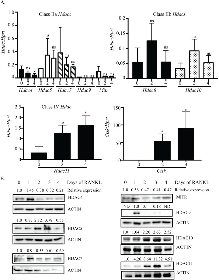
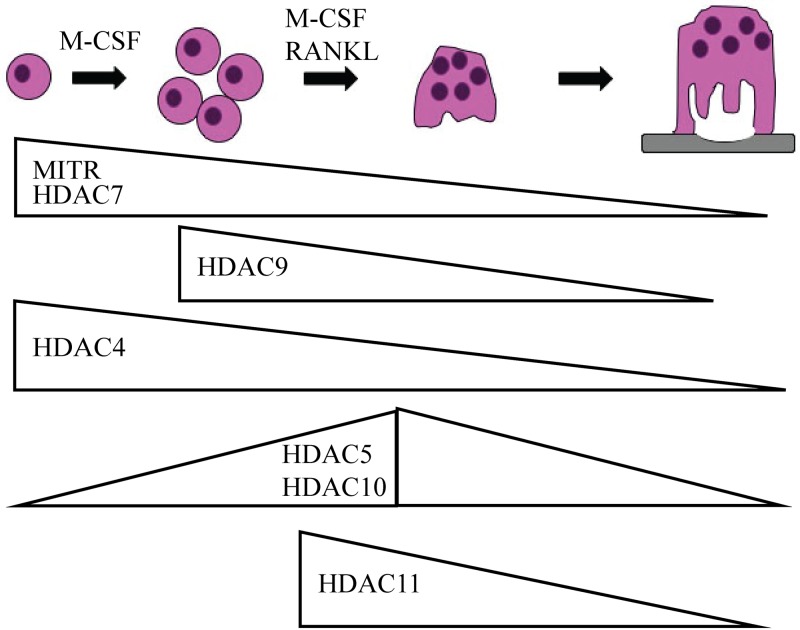
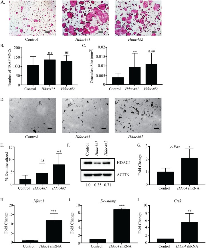
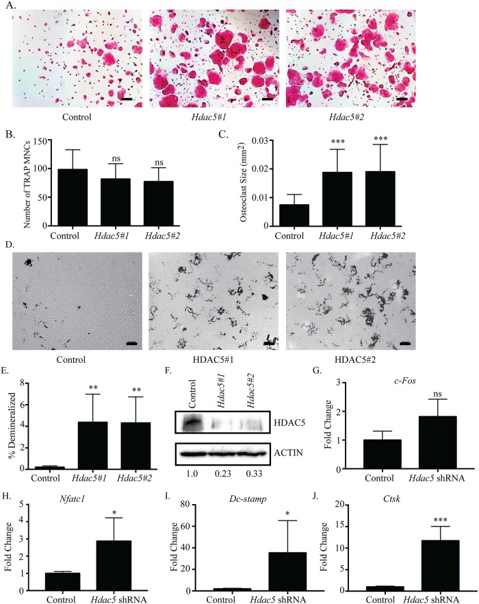
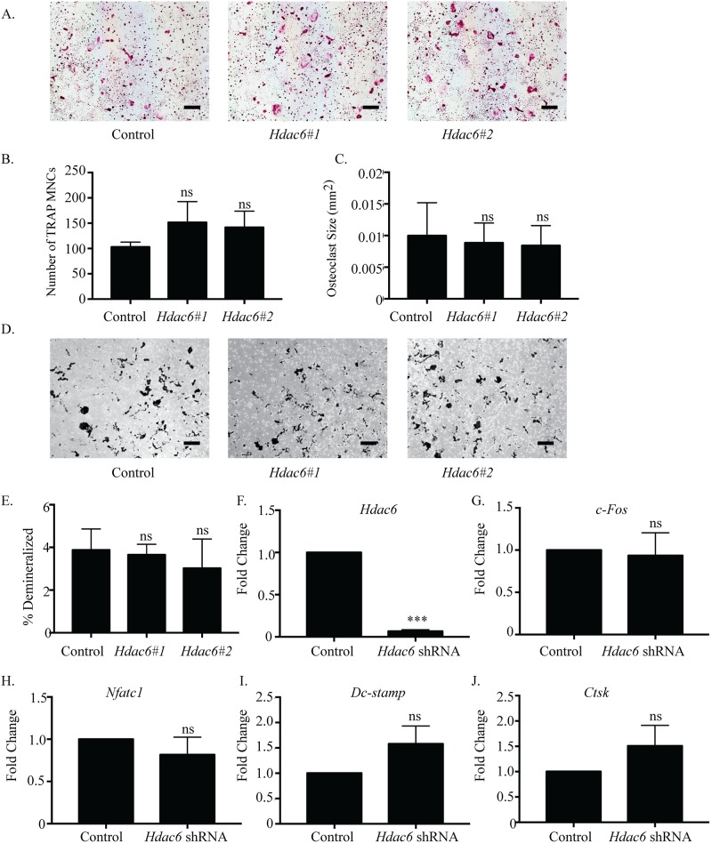
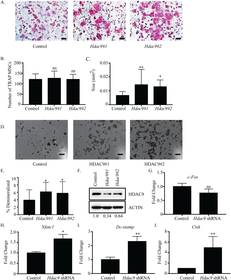
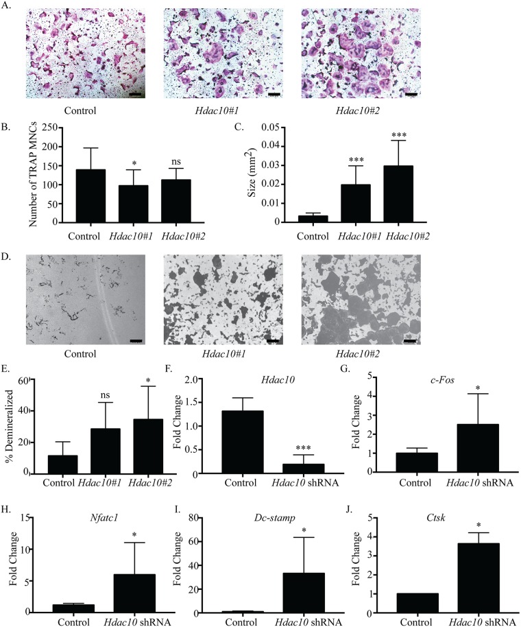
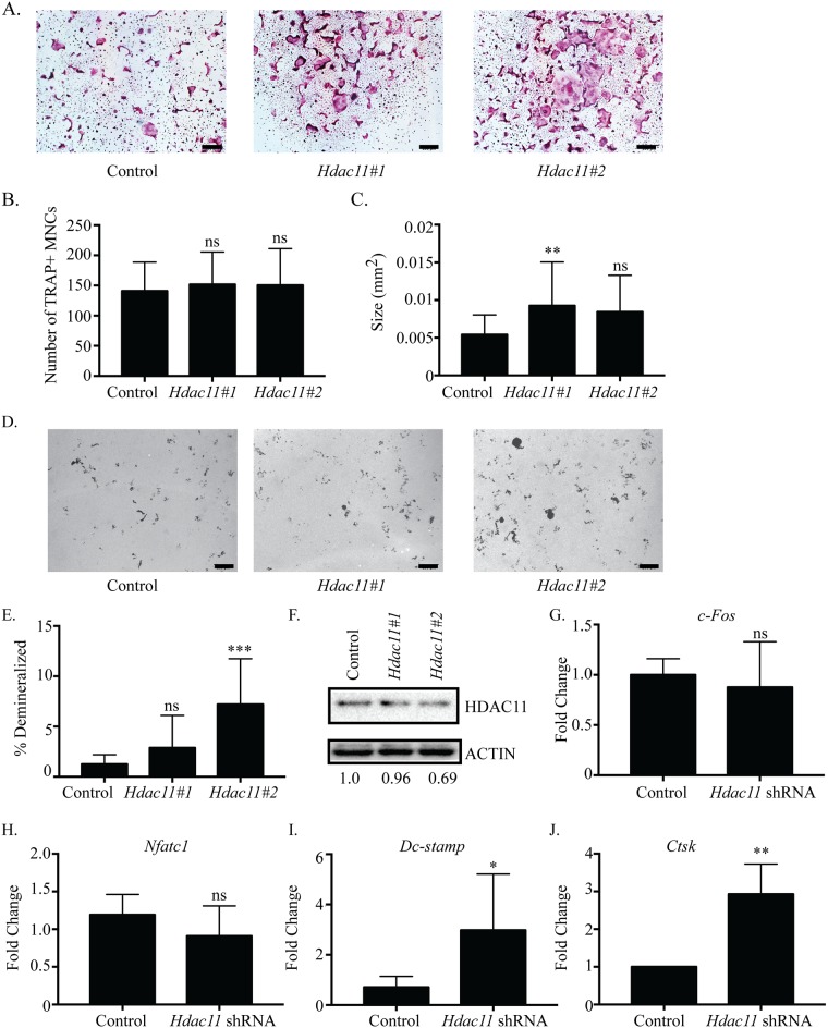
Histone deacetylases (HDACs) are negative regulators of transcription and have been shown to regulate specific changes in gene expression. In vertebrates, eighteen HDACs have thus far been identified and subdivided into four classes (I-IV). Key roles for several HDACs in bone development and biology have been elucidated through in vitro and in vivo models. By comparison, there is a paucity of data on the roles of individual HDACs in osteoclast formation and function. In this study, we investigated the gene expression patterns and the effects of suppressing individual class II (Hdac4, 5, 6, 9, and 10) and class IV (Hdac11) HDACs during osteoclast differentiation. We demonstrated that HDAC class II and IV members are differentially expressed during osteoclast differentiation. Additionally, individual shRNA-mediated suppression of Hdac4, 5, 9, 10 and 11 expression resulted in increased multinucleated osteoclast size and demineralization activity, with little to no change in the overall number of multinucleated osteoclasts formed compared with control shRNA-treated cells. We also detected increased expression of genes highly expressed in osteoclasts, including c-Fos, Nfatc1, Dc-stamp and Cathepsin K. These observations indicate that HDACs cooperatively regulate shared targets in a non-redundant manner.

摘要

组蛋白去乙酰化酶(HDACs)是转录的负调控因子,已被证明可调节基因表达的特定变化。在脊椎动物中,迄今为止已鉴定出18种HDACs,并将其分为四类(I-IV)。通过体外和体内模型已阐明了几种HDACs在骨骼发育和生物学中的关键作用。相比之下,关于单个HDACs在破骨细胞形成和功能中的作用的数据却很少。在本研究中,我们研究了破骨细胞分化过程中II类(Hdac4、5、6、9和10)和IV类(Hdac11)单个HDACs的基因表达模式及其抑制作用的影响。我们证明,HDAC II类和IV类成员在破骨细胞分化过程中差异表达。此外,单个shRNA介导的Hdac4、5、9、10和11表达抑制导致多核破骨细胞大小和脱矿活性增加,与对照shRNA处理的细胞相比,形成的多核破骨细胞总数几乎没有变化。我们还检测到破骨细胞中高表达的基因,包括c-Fos、Nfatc1、Dc-stamp和组织蛋白酶K的表达增加。这些观察结果表明,HDACs以非冗余方式协同调节共同的靶标。

https://cdn.ncbi.nlm.nih.gov/pmc/blobs/07c3/5617211/59e06caa5cdd/pone.0185441.g008.jpg
https://cdn.ncbi.nlm.nih.gov/pmc/blobs/07c3/5617211/95ab9f85e1f2/pone.0185441.g001.jpg
https://cdn.ncbi.nlm.nih.gov/pmc/blobs/07c3/5617211/9fb83087de8d/pone.0185441.g002.jpg
https://cdn.ncbi.nlm.nih.gov/pmc/blobs/07c3/5617211/dda09564da7d/pone.0185441.g003.jpg
https://cdn.ncbi.nlm.nih.gov/pmc/blobs/07c3/5617211/9dddb47cb634/pone.0185441.g004.jpg
https://cdn.ncbi.nlm.nih.gov/pmc/blobs/07c3/5617211/baa0ed8420eb/pone.0185441.g005.jpg
https://cdn.ncbi.nlm.nih.gov/pmc/blobs/07c3/5617211/2931b4299dcb/pone.0185441.g006.jpg
https://cdn.ncbi.nlm.nih.gov/pmc/blobs/07c3/5617211/f68bb26f0e00/pone.0185441.g007.jpg
https://cdn.ncbi.nlm.nih.gov/pmc/blobs/07c3/5617211/59e06caa5cdd/pone.0185441.g008.jpg

相似文献

1
Class II and IV HDACs function as inhibitors of osteoclast differentiation.II类和IV类组蛋白去乙酰化酶作为破骨细胞分化的抑制剂发挥作用。
PLoS One. 2017 Sep 27;12(9):e0185441. doi: 10.1371/journal.pone.0185441. eCollection 2017.
2
Inhibitors of histone deacetylases in class I and class II suppress human osteoclasts in vitro.I 类和 II 类组蛋白去乙酰化酶抑制剂可抑制体外人破骨细胞。
J Cell Physiol. 2011 Dec;226(12):3233-41. doi: 10.1002/jcp.22684.
3
HDAC3 and HDAC7 have opposite effects on osteoclast differentiation.HDAC3 和 HDAC7 对破骨细胞分化有相反的影响。
J Biol Chem. 2011 Apr 8;286(14):12056-65. doi: 10.1074/jbc.M110.216853. Epub 2011 Feb 15.
4
MS-275, a benzamide histone deacetylase inhibitor, prevents osteoclastogenesis by down-regulating c-Fos expression and suppresses bone loss in mice.MS-275,一种苯甲酰胺类组蛋白去乙酰化酶抑制剂,通过下调 c-Fos 表达来防止破骨细胞生成,并抑制小鼠的骨质流失。
Eur J Pharmacol. 2012 Sep 15;691(1-3):69-76. doi: 10.1016/j.ejphar.2012.07.034. Epub 2012 Jul 27.
5
Induction of DC-STAMP by alternative activation and downstream signaling mechanisms.通过交替激活和下游信号传导机制诱导DC-STAMP
J Bone Miner Res. 2007 Jul;22(7):992-1001. doi: 10.1359/jbmr.070401.
6
MiR-7b directly targets DC-STAMP causing suppression of NFATc1 and c-Fos signaling during osteoclast fusion and differentiation.微小RNA-7b直接靶向DC-STAMP,在破骨细胞融合与分化过程中导致核因子活化T细胞胞浆1和原癌基因c-Fos信号通路受到抑制。
Biochim Biophys Acta. 2014 Nov;1839(11):1084-96. doi: 10.1016/j.bbagrm.2014.08.002. Epub 2014 Aug 11.
7
Regulation of Osteoclast Differentiation and Skeletal Maintenance by Histone Deacetylases.组蛋白去乙酰化酶对破骨细胞分化和骨骼维持的调控。
Molecules. 2019 Apr 6;24(7):1355. doi: 10.3390/molecules24071355.
8
CTRP3 acts as a negative regulator of osteoclastogenesis through AMPK-c-Fos-NFATc1 signaling in vitro and RANKL-induced calvarial bone destruction in vivo.CTRP3在体外通过AMPK-c-Fos-NFATc1信号传导充当破骨细胞生成的负调节因子,并在体内抑制RANKL诱导的颅骨骨破坏。
Bone. 2015 Oct;79:242-51. doi: 10.1016/j.bone.2015.06.011. Epub 2015 Jun 21.
9
NFATc1 induces osteoclast fusion via up-regulation of Atp6v0d2 and the dendritic cell-specific transmembrane protein (DC-STAMP).NFATc1通过上调Atp6v0d2和树突状细胞特异性跨膜蛋白(DC-STAMP)诱导破骨细胞融合。
Mol Endocrinol. 2008 Jan;22(1):176-85. doi: 10.1210/me.2007-0237. Epub 2007 Sep 20.
10
Caffeine enhances osteoclast differentiation and maturation through p38 MAP kinase/Mitf and DC-STAMP/CtsK and TRAP pathway.咖啡因通过 p38 MAP 激酶/Mitf 和 DC-STAMP/CtsK 及 TRAP 通路增强破骨细胞分化和成熟。
Cell Signal. 2013 May;25(5):1222-7. doi: 10.1016/j.cellsig.2013.02.015. Epub 2013 Feb 19.

引用本文的文献

1
Identification of Critical Molecular Pathways Induced by HDAC11 Overexpression in Cardiac Mesenchymal Stem Cells.心脏间充质干细胞中HDAC11过表达诱导的关键分子途径的鉴定
Biomolecules. 2025 May 3;15(5):662. doi: 10.3390/biom15050662.
2
Network study of miRNA regulating traumatic heterotopic ossification.微小RNA调控创伤性异位骨化的网络研究
PLoS One. 2025 Feb 11;20(2):e0318779. doi: 10.1371/journal.pone.0318779. eCollection 2025.
3
Unlocking the Epigenetic Symphony: Histone Acetylation Orchestration in Bone Remodeling and Diseases.

本文引用的文献

1
Tubulin acetylation: responsible enzymes, biological functions and human diseases.微管蛋白乙酰化:相关酶、生物学功能及人类疾病
Cell Mol Life Sci. 2015 Nov;72(22):4237-55. doi: 10.1007/s00018-015-2000-5. Epub 2015 Jul 31.
2
Deletion of histone deacetylase 7 in osteoclasts decreases bone mass in mice by interactions with MITF.破骨细胞中组蛋白去乙酰化酶7的缺失通过与小眼畸形相关转录因子相互作用降低小鼠骨量。
PLoS One. 2015 Apr 15;10(4):e0123843. doi: 10.1371/journal.pone.0123843. eCollection 2015.
3
HDAC9 Inhibits Osteoclastogenesis via Mutual Suppression of PPARγ/RANKL Signaling.
解开表观遗传交响曲:骨重塑与疾病中的组蛋白乙酰化调控
Stem Cell Rev Rep. 2025 Feb;21(2):291-303. doi: 10.1007/s12015-024-10807-2. Epub 2024 Nov 4.
4
Roles of Histone Deacetylase 4 in the Inflammatory and Metabolic Processes.组蛋白去乙酰化酶 4 在炎症和代谢过程中的作用。
Diabetes Metab J. 2024 May;48(3):340-353. doi: 10.4093/dmj.2023.0174. Epub 2024 Mar 22.
5
Role and Regulation of Transcription Factors in Osteoclastogenesis.转录因子在破骨细胞分化中的作用与调控。
Int J Mol Sci. 2023 Nov 10;24(22):16175. doi: 10.3390/ijms242216175.
6
Targeting histone deacetylases for cancer therapy: Trends and challenges.以组蛋白去乙酰化酶为靶点进行癌症治疗:趋势与挑战。
Acta Pharm Sin B. 2023 Jun;13(6):2425-2463. doi: 10.1016/j.apsb.2023.02.007. Epub 2023 Feb 18.
7
Histone Deacetylase 6 Inhibitor CKD-WID Suppressed Monosodium Urate-Induced Osteoclast Formation by Blocking Calcineurin-NFAT Pathway in RAW 264.7 Cells.组蛋白去乙酰化酶6抑制剂CKD-WID通过阻断RAW 264.7细胞中的钙调神经磷酸酶-NFAT途径抑制尿酸钠诱导的破骨细胞形成。
Pharmaceuticals (Basel). 2023 Mar 16;16(3):446. doi: 10.3390/ph16030446.
8
FTY720 Attenuates LPS-Induced Inflammatory Bone Loss by Inhibiting Osteoclastogenesis via the NF-B and HDAC4/ATF Pathways.FTY720 通过抑制 NF-B 和 HDAC4/ATF 通路来抑制破骨细胞生成,从而减轻 LPS 诱导的炎症性骨丢失。
J Immunol Res. 2023 Jan 6;2023:8571649. doi: 10.1155/2023/8571649. eCollection 2023.
9
HDAC11, an emerging therapeutic target for metabolic disorders.HDAC11,代谢紊乱的新兴治疗靶点。
Front Endocrinol (Lausanne). 2022 Oct 20;13:989305. doi: 10.3389/fendo.2022.989305. eCollection 2022.
10
Histone deacetylase 5 is a phosphorylation substrate of protein kinase D in osteoclasts.组蛋白去乙酰化酶5是破骨细胞中蛋白激酶D的磷酸化底物。
Bone. 2022 Jun;159:116393. doi: 10.1016/j.bone.2022.116393. Epub 2022 Mar 19.
组蛋白去乙酰化酶9通过相互抑制PPARγ/RANKL信号通路抑制破骨细胞生成。
Mol Endocrinol. 2015 May;29(5):730-8. doi: 10.1210/me.2014-1365. Epub 2015 Mar 20.
4
Divergent roles of histone deacetylase 6 (HDAC6) and histone deacetylase 11 (HDAC11) on the transcriptional regulation of IL10 in antigen presenting cells.组蛋白去乙酰化酶 6(HDAC6)和组蛋白去乙酰化酶 11(HDAC11)在抗原提呈细胞中对 IL10 的转录调控中的差异作用。
Mol Immunol. 2014 Jul;60(1):44-53. doi: 10.1016/j.molimm.2014.02.019. Epub 2014 Apr 18.
5
Microtubule dynamic instability controls podosome patterning in osteoclasts through EB1, cortactin, and Src.微管动态不稳定性通过 EB1、桩蛋白和Src 控制破骨细胞中的破骨细胞足形成。
Mol Cell Biol. 2014 Jan;34(1):16-29. doi: 10.1128/MCB.00578-13. Epub 2013 Oct 21.
6
Inhibition of HDAC6 deacetylase activity increases its binding with microtubules and suppresses microtubule dynamic instability in MCF-7 cells.组蛋白去乙酰化酶 6(HDAC6)去乙酰化酶活性的抑制增加了其与微管的结合,并抑制 MCF-7 细胞中的微管动态不稳定性。
J Biol Chem. 2013 Aug 2;288(31):22516-26. doi: 10.1074/jbc.M113.489328. Epub 2013 Jun 24.
7
HDAC7 inhibits osteoclastogenesis by reversing RANKL-triggered β-catenin switch.组蛋白去乙酰化酶7通过逆转RANKL触发的β-连环蛋白转换来抑制破骨细胞生成。
Mol Endocrinol. 2013 Feb;27(2):325-35. doi: 10.1210/me.2012-1302. Epub 2012 Nov 30.
8
Modulation of histone deacetylase 6 (HDAC6) nuclear import and tubulin deacetylase activity through acetylation.通过乙酰化调节组蛋白去乙酰化酶 6(HDAC6)的核输入和微管蛋白去乙酰化酶活性。
J Biol Chem. 2012 Aug 17;287(34):29168-74. doi: 10.1074/jbc.M112.371120. Epub 2012 Jul 9.
9
Differential distribution of posttranslationally modified microtubules in osteoclasts.破骨细胞中翻译后修饰的微管的差异分布。
J Histochem Cytochem. 2011 Jun;59(6):630-8. doi: 10.1369/0022155411405334. Epub 2011 Mar 18.
10
RANKL induces NFATc1 acetylation and stability via histone acetyltransferases during osteoclast differentiation.核因子活化 T 细胞受体相关因子(NFATc1)在破骨细胞分化过程中,通过组蛋白乙酰转移酶诱导其乙酰化和稳定性。
Biochem J. 2011 Jun 1;436(2):253-62. doi: 10.1042/BJ20110062.