Magat Guldane, Ozcan Sevgi
Department of Oral Radiology Faculty of Dentistry Necmettin Erbakan University Turkey.
J Istanb Univ Fac Dent. 2017 Apr 3;51(2):29-36. doi: 10.17096/jiufd.35768. eCollection 2017.
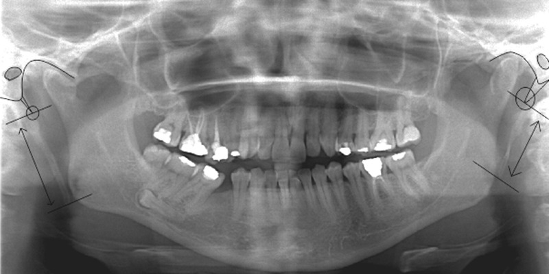
The purpose of this study was to investigate the morphology and calcification pattern of the styloid process (SP) and to determine their relations with subjects' age, gender, and dental status.
910 panoramic radiographs were stratified by age, dental status and gender. The distance between the points where SP leaves the tympanic plate of the temporal bone and the bony tip of SP was measured. Calcification patterns were classified as : (A) Region 1, tympanohyal alone (B) Region 2, stylohyal alone (C) Region 1 and 2, separate (D) Regions 1 and 2, continuous (E) Regions 1, 2, and 3, continuous (F) Regions 1, 2, and 3, separate (G) Regions 1 and 2, continuous, but separate from 3 (H) Regions 2 and 3, separate (I) Regions 2 and 3, continuous, but separate from 1 (J) Region 3 alone (K) Region 3 and 4, continuous (may include calcification in one other region) (L) No styloid process visible.
The right SPs were found to be longer than the left (p<0.05). Types D (right 42.9%, left 42%) and E (right 33.3%, left 30.8%) were the most common morphological calcifications on both sides. No statistical difference was found for bilateral SP length between gender, age, and dental status groups. A significant difference was found only for right SP morphological calcification types as to age groups in both genders (p<0.05). No significant difference was found for SP morphological calcification types according to gender and dental status.
The morphological types are formed at their present area. Even though SP calcification type was determined according to the length of SP, age was not an effective factor on the length, but the morphological calcification type of SP. Therefore, factors other than age may have a role in the development of morphological calcification types. Structural characteristics of SP are not associated with age, gender and dental status.
本研究旨在探讨茎突(SP)的形态和钙化模式,并确定它们与受试者年龄、性别及牙齿状况的关系。
910张全景X线片按年龄、牙齿状况和性别进行分层。测量茎突离开颞骨鼓板处与茎突骨尖之间的距离。钙化模式分为:(A)区域1,仅鼓室舌骨;(B)区域2,仅茎突舌骨;(C)区域1和2,分离;(D)区域1和2,连续;(E)区域1、2和3,连续;(F)区域1、2和3,分离;(G)区域1和2,连续,但与区域3分离;(H)区域2和3,分离;(I)区域2和3,连续,但与区域1分离;(J)仅区域3;(K)区域3和4,连续(可能包括其他一个区域的钙化);(L)未见茎突。
发现右侧茎突比左侧长(p<0.05)。类型D(右侧42.9%,左侧42%)和E(右侧33.3%,左侧30.8%)是两侧最常见的形态学钙化类型。在性别、年龄和牙齿状况组之间,双侧茎突长度未发现统计学差异。仅在两性的年龄组中,右侧茎突形态学钙化类型存在显著差异(p<0.05)。根据性别和牙齿状况,茎突形态学钙化类型未发现显著差异。
形态学类型在其当前区域形成。尽管茎突钙化类型是根据茎突长度确定的,但年龄不是影响茎突长度的有效因素,而是影响茎突形态学钙化类型的因素。因此,除年龄外的其他因素可能在形态学钙化类型的形成中起作用。茎突的结构特征与年龄、性别和牙齿状况无关。