• 文献检索
  • 文档翻译
  • 深度研究
  • 学术资讯
  • Suppr Zotero 插件Zotero 插件
  • 邀请有礼
  • 套餐&价格
  • 历史记录
应用&插件
Suppr Zotero 插件Zotero 插件浏览器插件Mac 客户端Windows 客户端微信小程序
定价
高级版会员购买积分包购买API积分包
服务
文献检索文档翻译深度研究API 文档MCP 服务
关于我们
关于 Suppr公司介绍联系我们用户协议隐私条款
关注我们

Suppr 超能文献

核心技术专利:CN118964589B侵权必究
粤ICP备2023148730 号-1Suppr @ 2026

文献检索

告别复杂PubMed语法,用中文像聊天一样搜索,搜遍4000万医学文献。AI智能推荐,让科研检索更轻松。

立即免费搜索

文件翻译

保留排版,准确专业,支持PDF/Word/PPT等文件格式,支持 12+语言互译。

免费翻译文档

深度研究

AI帮你快速写综述,25分钟生成高质量综述,智能提取关键信息,辅助科研写作。

立即免费体验

癌症“种子”在腹膜的“土壤”里:腹腔内癌症转移发病机制的全面综述。

The peritoneal "soil" for a cancerous "seed": a comprehensive review of the pathogenesis of intraperitoneal cancer metastases.

机构信息

Department of Hypertensiology, Angiology and Internal Medicine, Poznań University of Medical Sciences, Długa 1/2 Str., 61-848, Poznan, Poland.

出版信息

Cell Mol Life Sci. 2018 Feb;75(3):509-525. doi: 10.1007/s00018-017-2663-1. Epub 2017 Sep 27.

DOI:10.1007/s00018-017-2663-1
PMID:28956065
原文链接:https://pmc.ncbi.nlm.nih.gov/articles/PMC5765197/
Abstract

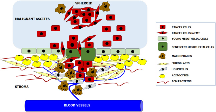
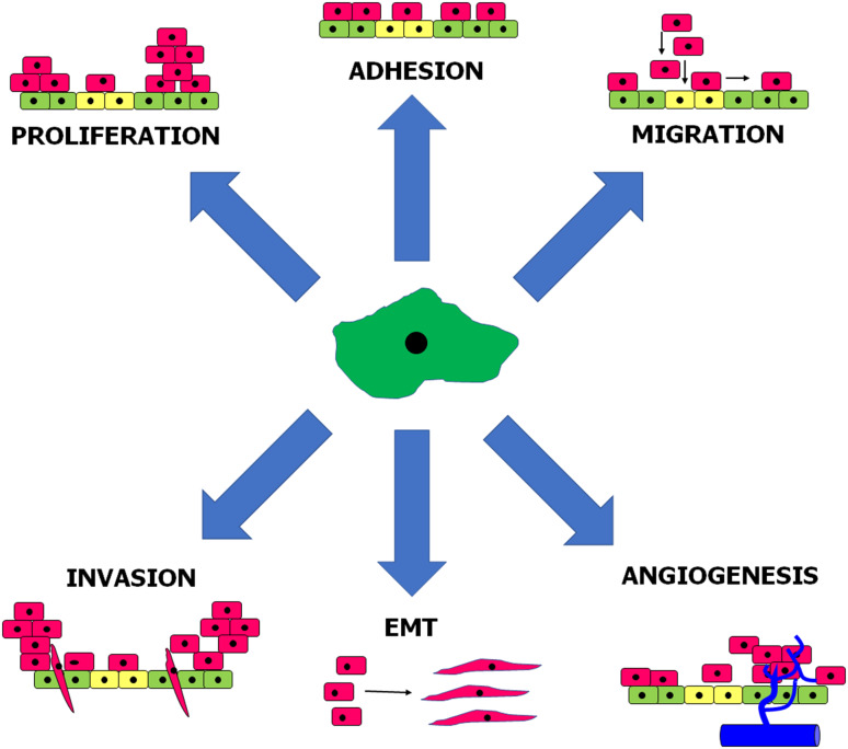
Various types of tumors, particularly those originating from the ovary and gastrointestinal tract, display a strong predilection for the peritoneal cavity as the site of metastasis. The intraperitoneal spread of a malignancy is orchestrated by a reciprocal interplay between invading cancer cells and resident normal peritoneal cells. In this review, we address the current state-of-art regarding colonization of the peritoneal cavity by ovarian, colorectal, pancreatic, and gastric tumors. Particular attention is paid to the pro-tumoral role of various kinds of peritoneal cells, including mesothelial cells, fibroblasts, adipocytes, macrophages, the vascular endothelium, and hospicells. Anatomo-histological considerations on the pro-metastatic environment of the peritoneal cavity are presented in the broader context of organ-specific development of distal metastases in accordance with Paget's "seed and soil" theory of tumorigenesis. The activity of normal peritoneal cells during pivotal elements of cancer progression, i.e., adhesion, migration, invasion, proliferation, EMT, and angiogenesis, is discussed from the perspective of well-defined general knowledge on a hospitable tumor microenvironment created by the cellular elements of reactive stroma, such as cancer-associated fibroblasts and macrophages. Finally, the paper addresses the unique features of the peritoneal cavity that predispose this body compartment to be a niche for cancer metastases, presents issues that are topics of an ongoing debate, and points to areas that still require further in-depth investigations.

摘要

各种类型的肿瘤,特别是源自卵巢和胃肠道的肿瘤,强烈倾向于将腹膜腔作为转移部位。恶性肿瘤的腹膜内扩散是由侵袭性癌细胞和驻留的正常腹膜细胞之间的相互作用协调的。在这篇综述中,我们讨论了卵巢癌、结直肠癌、胰腺癌和胃癌在腹膜腔内定植的最新进展。特别关注各种腹膜细胞,包括间皮细胞、成纤维细胞、脂肪细胞、巨噬细胞、血管内皮细胞和休眠细胞,对肿瘤的促瘤作用。根据 Paget 的肿瘤发生“种子与土壤”理论,在腹膜腔中有利于转移的环境的解剖组织学考虑,是在与器官特异性远处转移相关的更广泛背景下提出的。从反应性基质的细胞成分(如癌相关成纤维细胞和巨噬细胞)所创建的有利于肿瘤的微环境的明确的一般知识的角度,讨论了正常腹膜细胞在癌症进展的关键环节(即黏附、迁移、侵袭、增殖、EMT 和血管生成)中的活性。本文还讨论了腹膜腔的独特特征,这些特征使该体腔容易成为癌症转移的龛位,提出了正在讨论的问题,并指出了仍需要进一步深入研究的领域。

https://cdn.ncbi.nlm.nih.gov/pmc/blobs/32a6/11105450/6a12251042b2/18_2017_2663_Fig2_HTML.jpg
https://cdn.ncbi.nlm.nih.gov/pmc/blobs/32a6/11105450/7dd5fae8e289/18_2017_2663_Fig1_HTML.jpg
https://cdn.ncbi.nlm.nih.gov/pmc/blobs/32a6/11105450/6a12251042b2/18_2017_2663_Fig2_HTML.jpg
https://cdn.ncbi.nlm.nih.gov/pmc/blobs/32a6/11105450/7dd5fae8e289/18_2017_2663_Fig1_HTML.jpg
https://cdn.ncbi.nlm.nih.gov/pmc/blobs/32a6/11105450/6a12251042b2/18_2017_2663_Fig2_HTML.jpg

相似文献

1
The peritoneal "soil" for a cancerous "seed": a comprehensive review of the pathogenesis of intraperitoneal cancer metastases.癌症“种子”在腹膜的“土壤”里:腹腔内癌症转移发病机制的全面综述。
Cell Mol Life Sci. 2018 Feb;75(3):509-525. doi: 10.1007/s00018-017-2663-1. Epub 2017 Sep 27.
2
Procancerogenic activity of senescent cells: A case of the peritoneal mesothelium.衰老细胞的致癌活性:以腹膜间皮为例。
Ageing Res Rev. 2018 May;43:1-9. doi: 10.1016/j.arr.2018.01.002. Epub 2018 Jan 31.
3
Intraperitoneal metastasis of ovarian cancer: new insights on resident macrophages in the peritoneal cavity.卵巢癌腹腔转移:腹腔固有巨噬细胞的新见解。
Front Immunol. 2023 Apr 25;14:1104694. doi: 10.3389/fimmu.2023.1104694. eCollection 2023.
4
Mesothelial cells differentiate into fibroblast-like cells under the scirrhous gastric cancer microenvironment and promote peritoneal carcinomatosis in vitro and in vivo.间皮细胞在硬癌胃癌微环境下向成纤维细胞样细胞分化,并在体外和体内促进腹膜癌转移。
Mol Cell Biochem. 2013 May;377(1-2):177-85. doi: 10.1007/s11010-013-1583-0. Epub 2013 Feb 8.
5
CD90+ mesothelial-like cells in peritoneal fluid promote peritoneal metastasis by forming a tumor permissive microenvironment.腹腔液中 CD90+间皮样细胞通过形成肿瘤许可的微环境促进腹膜转移。
PLoS One. 2014 Jan 21;9(1):e86516. doi: 10.1371/journal.pone.0086516. eCollection 2014.
6
Carcinoma-associated fibroblasts derive from mesothelial cells via mesothelial-to-mesenchymal transition in peritoneal metastasis.癌相关成纤维细胞通过腹膜转移中的间皮细胞向间充质转化而来。
J Pathol. 2013 Dec;231(4):517-31. doi: 10.1002/path.4281.
7
Senescent peritoneal mesothelium creates a niche for ovarian cancer metastases.衰老的腹膜间皮为卵巢癌转移创造了一个微环境。
Cell Death Dis. 2016 Dec 29;7(12):e2565. doi: 10.1038/cddis.2016.417.
8
Development of Peritoneal Carcinomatosis in Epithelial Ovarian Cancer: A Review.上皮性卵巢癌腹膜转移的发生机制:综述
J Histochem Cytochem. 2018 Feb;66(2):67-83. doi: 10.1369/0022155417742897. Epub 2017 Nov 22.
9
Mesothelial-to-Mesenchymal Transition and Exosomes in Peritoneal Metastasis of Ovarian Cancer.间皮细胞向间充质转化与卵巢癌腹膜转移中的外泌体。
Int J Mol Sci. 2021 Oct 25;22(21):11496. doi: 10.3390/ijms222111496.
10
Host Wnt5a Potentiates Microenvironmental Regulation of Ovarian Cancer Metastasis.宿主 Wnt5a 增强卵巢癌细胞转移的微环境调控。
Cancer Res. 2020 Mar 1;80(5):1156-1170. doi: 10.1158/0008-5472.CAN-19-1601. Epub 2020 Jan 13.

引用本文的文献

1
Prognostic value of multiparameter [Ga]Ga-DOTA-FAPI-04 PET/MR imaging biomarkers for patients with advanced pancreatic cancer.多参数[镓]镓-DOTA-FAPI-04 PET/MR成像生物标志物对晚期胰腺癌患者的预后价值
Eur J Nucl Med Mol Imaging. 2025 Sep 9. doi: 10.1007/s00259-025-07491-w.
2
Regulatory B cells drive immune evasion in the tumor microenvironment and are involved peritoneal metastasis in gastric cancer.调节性B细胞在肿瘤微环境中驱动免疫逃逸,并参与胃癌的腹膜转移。
Sci Rep. 2025 Jul 28;15(1):27499. doi: 10.1038/s41598-025-11887-x.
3
CAMKKβ supports growth and viability of epithelial ovarian cancer in vitro and in vivo.

本文引用的文献

1
Diverse properties of the mesothelial cells in health and disease.健康与疾病状态下间皮细胞的多种特性。
Pleura Peritoneum. 2016 Jun 1;1(2):79-89. doi: 10.1515/pp-2016-0009. Epub 2016 Jun 9.
2
The protective activity of mesothelial cells against peritoneal growth of gastrointestinal tumors: The role of soluble ICAM-1.间皮细胞对胃肠道肿瘤腹膜种植的保护作用:可溶性细胞间黏附分子-1的作用
Int J Biochem Cell Biol. 2017 May;86:26-31. doi: 10.1016/j.biocel.2017.03.013. Epub 2017 Mar 18.
3
Chronic inflammation confers to the metabolic reprogramming associated with tumorigenesis of colorectal cancer.
钙调神经磷酸酶激酶β(CAMKKβ)在体外和体内均支持上皮性卵巢癌的生长和存活。
Sci Rep. 2025 Jul 17;15(1):25913. doi: 10.1038/s41598-025-11584-9.
4
Conversion Surgery After Trastuzumab-Based Chemotherapy and Immunotherapy for Stage IV HER2-Positive Gastric Cancer: A Retrospective Multicenter Cohort Study.曲妥珠单抗为基础的化疗和免疫治疗后IV期HER2阳性胃癌的转化手术:一项回顾性多中心队列研究
Ann Surg Oncol. 2025 Jun 26. doi: 10.1245/s10434-025-17729-4.
5
The Tumor-Associated Neutrophils-Related Signatures Predict Prognosis and Indicate Immune Landscape in Colorectal Carcinoma.肿瘤相关中性粒细胞相关特征预测结直肠癌预后并指示免疫格局
Mediators Inflamm. 2025 Jun 5;2025:7259278. doi: 10.1155/mi/7259278. eCollection 2025.
6
Intraoperatively preventive intraperitoneal perfusion chemotherapy with lobaplatin in colorectal cancer: a prospective, randomised, controlled, multicentre study.术中腹腔内预防性灌注洛铂用于结直肠癌治疗:一项前瞻性、随机、对照、多中心研究
BMC Med. 2025 Jun 6;23(1):336. doi: 10.1186/s12916-025-04180-1.
7
Characteristics of F-FAPI-04 PET/CT in patients with peritoneal metastasis and to predict treatment efficacy, a head-to-head comparison with F-FDG PET/CT.F-FAPI-04 PET/CT在腹膜转移患者中的特征及预测治疗疗效:与F-FDG PET/CT的直接比较
Cancer Imaging. 2025 Jun 2;25(1):66. doi: 10.1186/s40644-025-00887-9.
8
Imaging gastric cancer metastasis progression in an organotypic, three-dimensional functional model of the human peritoneum.在人腹膜的器官型三维功能模型中对胃癌转移进展进行成像。
Pleura Peritoneum. 2025 Mar 6;10(1):11-17. doi: 10.1515/pp-2024-0020. eCollection 2025 Mar.
9
Comparison of the diagnostic value of [Ga]Ga-FAP-2286 PET/CT and [F]-FDG PET/CT imaging in different types of pleural and peritoneal metastatic tumors.[镓]Ga-FAP-2286 PET/CT与[氟]F-FDG PET/CT成像在不同类型胸膜和腹膜转移性肿瘤中的诊断价值比较。
Eur J Nucl Med Mol Imaging. 2025 Apr 24. doi: 10.1007/s00259-025-07265-4.
10
Paired primary-metastasis patient-derived organoids and mouse models identify phenotypic evolution and druggable dependencies of peritoneal metastasis from appendiceal cancer.配对的原发性-转移性患者来源类器官和小鼠模型确定了阑尾癌腹膜转移的表型演变和可靶向依赖性。
bioRxiv. 2025 Feb 22:2025.02.17.638725. doi: 10.1101/2025.02.17.638725.
慢性炎症赋予与结直肠癌肿瘤发生相关的代谢重编程。
Cancer Biol Ther. 2017 Apr 3;18(4):237-244. doi: 10.1080/15384047.2017.1294292. Epub 2017 Feb 25.
4
Malignant ascites-derived exosomes promote proliferation and induce carcinoma-associated fibroblasts transition in peritoneal mesothelial cells.恶性腹水来源的外泌体促进腹膜间皮细胞增殖并诱导癌相关成纤维细胞转变。
Oncotarget. 2017 Jun 27;8(26):42262-42271. doi: 10.18632/oncotarget.15040.
5
A loop of cancer-stroma-cancer interaction promotes peritoneal metastasis of ovarian cancer via TNFα-TGFα-EGFR.癌-基质-癌相互作用循环通过TNFα-TGFα-EGFR促进卵巢癌的腹膜转移。
Oncogene. 2017 Jun 22;36(25):3576-3587. doi: 10.1038/onc.2016.509. Epub 2017 Feb 6.
6
Senescent peritoneal mesothelium creates a niche for ovarian cancer metastases.衰老的腹膜间皮为卵巢癌转移创造了一个微环境。
Cell Death Dis. 2016 Dec 29;7(12):e2565. doi: 10.1038/cddis.2016.417.
7
Obesity and cancer: the role of adipose tissue and adipo-cytokines-induced chronic inflammation.肥胖与癌症:脂肪组织及脂肪细胞因子诱导的慢性炎症所起的作用
J Cancer. 2016 Nov 26;7(15):2346-2359. doi: 10.7150/jca.16884. eCollection 2016.
8
Omental adipocytes enhance the invasiveness of gastric cancer cells by oleic acid-induced activation of the PI3K-Akt signaling pathway.网膜脂肪细胞通过油酸诱导的PI3K-Akt信号通路激活增强胃癌细胞的侵袭性。
Int J Biochem Cell Biol. 2017 Mar;84:14-21. doi: 10.1016/j.biocel.2016.12.002. Epub 2016 Dec 9.
9
Exosomes Promote Ovarian Cancer Cell Invasion through Transfer of CD44 to Peritoneal Mesothelial Cells.外泌体通过将 CD44 转移至腹膜间皮细胞促进卵巢癌细胞侵袭。
Mol Cancer Res. 2017 Jan;15(1):78-92. doi: 10.1158/1541-7786.MCR-16-0191. Epub 2016 Oct 6.
10
Tumor-associated macrophages drive spheroid formation during early transcoelomic metastasis of ovarian cancer.肿瘤相关巨噬细胞在卵巢癌早期经体腔转移过程中驱动球体形成。
J Clin Invest. 2016 Nov 1;126(11):4157-4173. doi: 10.1172/JCI87252. Epub 2016 Oct 10.