Beach Paul A, Huck Jonathan T, Zhu David C, Bozoki Andrea C
D.O., Ph.D. Training Program, Michigan State University College of Osteopathic MedicineEast Lansing, MI, United States.
Neuroscience Program, Michigan State UniversityEast Lansing, MI, United States.
Front Aging Neurosci. 2017 Sep 14;9:297. doi: 10.3389/fnagi.2017.00297. eCollection 2017.
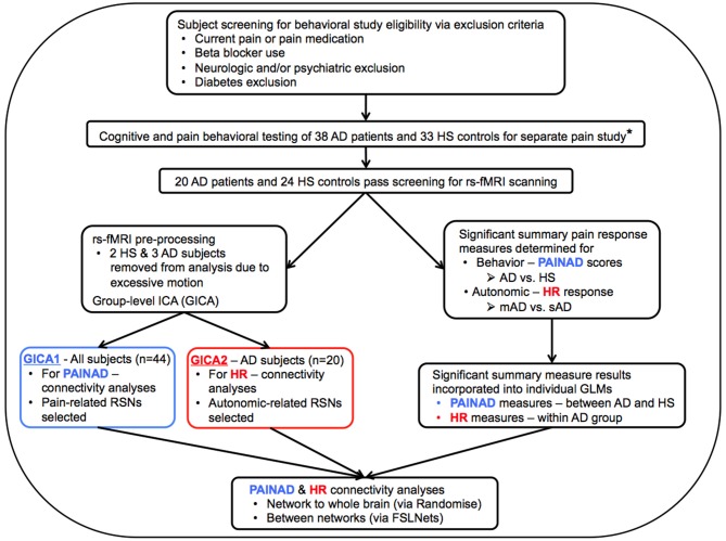
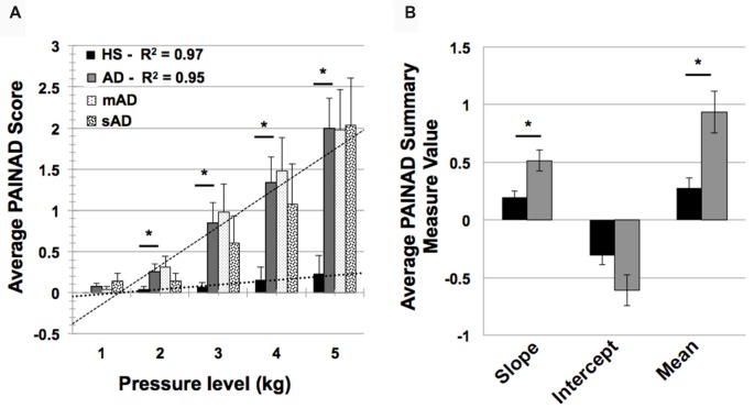
While pain behaviors are increased in Alzheimer's disease (AD) patients compared to healthy seniors (HS) across multiple disease stages, autonomic responses are reduced with advancing AD. To better understand the neural mechanisms underlying these phenomena, we undertook a controlled cross-sectional study examining behavioral (Pain Assessment in Advanced Dementia, PAINAD scores) and autonomic (heart rate, HR) pain responses in 24 HS and 20 AD subjects using acute pressure stimuli. Resting-state fMRI was utilized to investigate how group connectivity differences were related to altered pain responses. Pain behaviors (slope of PAINAD score change and mean PAINAD score) were increased in patients vs.
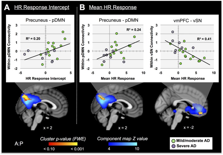
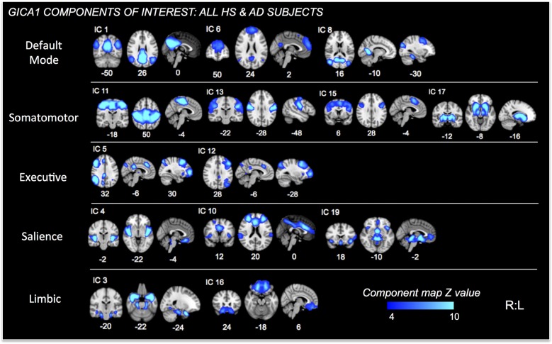
Autonomic measures (HR change intercept and mean HR change) were reduced in severe vs. mildly affected AD patients. Group functional connectivity differences associated with greater pain behavior reactivity in patients included: connectivity within a temporal limbic network (TLN) and between the TLN and ventromedial prefrontal cortex (vmPFC); between default mode network (DMN) subcomponents; between the DMN and ventral salience network (vSN). Reduced HR responses within the AD group were associated with connectivity changes within the DMN and vSN-specifically the precuneus and vmPFC. Discriminant classification indicated HR-related connectivity within the vSN to the vmPFC best distinguished AD severity. Thus, altered behavioral and autonomic pain responses in AD reflects dysfunction of networks and structures subserving affective, self-reflective, salience and autonomic regulation.
与健康老年人(HS)相比,阿尔茨海默病(AD)患者在多个疾病阶段的疼痛行为均有所增加,但随着AD病情进展,自主神经反应会减弱。为了更好地理解这些现象背后的神经机制,我们进行了一项对照横断面研究,使用急性压力刺激检查了24名HS和20名AD受试者的行为(晚期痴呆疼痛评估,PAINAD评分)和自主神经(心率,HR)疼痛反应。利用静息态功能磁共振成像来研究组间连接差异与疼痛反应改变之间的关系。与对照组相比,患者的疼痛行为(PAINAD评分变化斜率和平均PAINAD评分)增加。
与轻度受影响的AD患者相比,重度AD患者的自主神经测量指标(HR变化截距和平均HR变化)降低。与患者更大的疼痛行为反应性相关的组功能连接差异包括:颞叶边缘网络(TLN)内以及TLN与腹内侧前额叶皮层(vmPFC)之间的连接;默认模式网络(DMN)子成分之间的连接;DMN与腹侧突显网络(vSN)之间的连接。AD组内HR反应降低与DMN和vSN内的连接变化有关,特别是楔前叶和vmPFC。判别分类表明,vSN内与vmPFC的HR相关连接最能区分AD的严重程度。因此,AD中行为和自主神经疼痛反应的改变反映了服务于情感、自我反思、突显和自主调节的网络和结构的功能障碍。