Piehowski Paul D, Zhao Rui, Moore Ronald J, Clair Geremy, Ansong Charles
Biological Sciences Division, Pacific Northwest National Laboratory, Richland, WA, USA.
Environmental Molecular Sciences Laboratory, Pacific Northwest National Laboratory, Richland, WA, USA.
Methods Mol Biol. 2018;1788:269-277. doi: 10.1007/7651_2017_78.
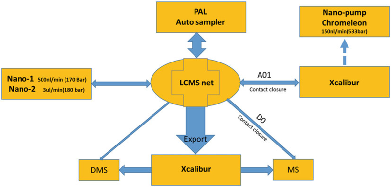
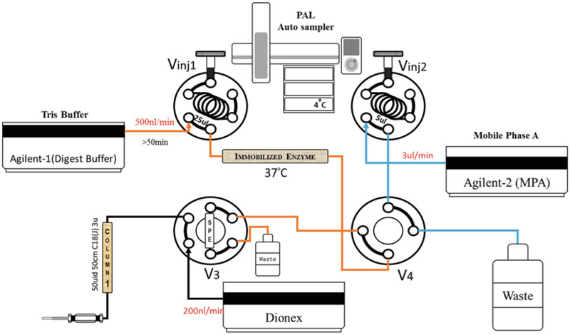
Traditionally, proteomic studies have been carried out on whole tissues or organs enabling the profiling of thousands of proteins within a single LC-MS analysis. A disadvantage of this approach is that proteomes generated from whole tissues are an "average" that represents a blend of cell types and distinct anatomical regions which can obscure important biological phenomena. Laser capture microdissection (LCM) is an elegant method that allows tissue features of interest, as small as a single cell, to be identified and isolated for downstream analysis. Herein we describe an approach that utilizes an immobilized enzyme reactor (IMER) coupled directly to nanoLC-MS/MS for highly sensitive, automated, quantitative proteomic analysis of the microscopic tissue specimens generated by LCM.
传统上,蛋白质组学研究是在整个组织或器官上进行的,能够在一次液相色谱-质谱分析中对数千种蛋白质进行分析。这种方法的一个缺点是,从整个组织生成的蛋白质组是一种“平均值”,它代表了细胞类型和不同解剖区域的混合,这可能会掩盖重要的生物学现象。激光捕获显微切割(LCM)是一种精妙的方法,它可以识别并分离出感兴趣的组织特征,小到单个细胞,以便进行下游分析。在此,我们描述了一种方法,该方法利用直接与纳升液相色谱-串联质谱联用的固定化酶反应器(IMER),对LCM生成的微观组织标本进行高灵敏度、自动化的定量蛋白质组学分析。