Department of Biological Science and Technology, China Medical University, Taichung 40402, Taiwan, R.O.C.
Department of Food Nutrition and Health Biotechnology, Asia University, Taichung 41354, Taiwan, R.O.C.
Mol Med Rep. 2017 Dec;16(6):7959-7966. doi: 10.3892/mmr.2017.7651. Epub 2017 Sep 28.
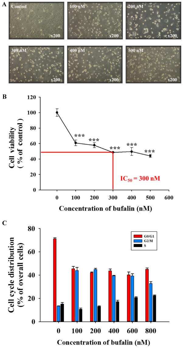
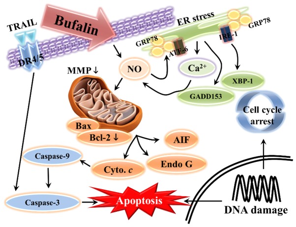
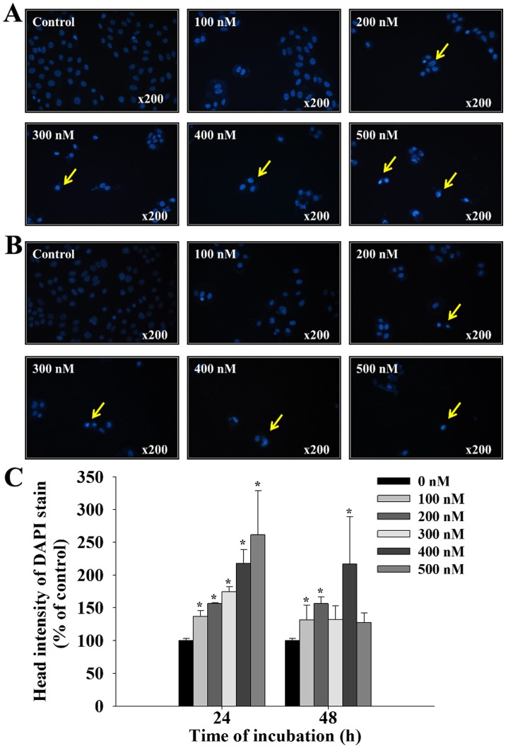
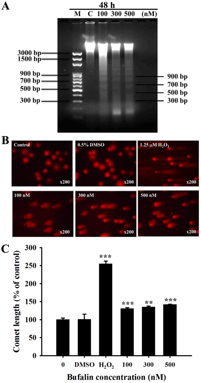
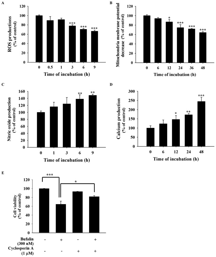
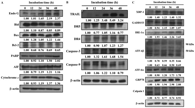
The aim of the present study was to investigate the cytotoxic effects of bufalin on SCC‑4 human tongue cancer cells. Cell morphological changes and viability were examined using phase contrast microscopy and flow cytometry, respectively. The results indicated that bufalin induced morphological changes and reduced total viable cells. Apoptotic cell death was analyzed by DAPI staining and DNA gel electrophoresis; the results revealed that bufalin induced cell apoptosis. Levels of reactive oxygen species (ROS), Ca2+, nitric oxide (NO) and mitochondrial membrane potential (ΔΨm) were measured by flow cytometry, and bufalin was observed to increase Ca2+ and NO production, decrease the ΔΨm and reduce ROS production in SCC‑4 cells. In addition, western blotting was performed to detect apoptosis‑associated protein expression. The results demonstrated that bufalin reduced the expression of the anti‑apoptotic protein B‑cell lymphoma 2 (Bcl‑2) and increased the expression of the pro‑apoptotic protein, Bcl‑2‑associated X protein. However, bufalin treatment also increased the expression of other apoptosis‑associated proteins such as apoptosis‑inducing factor and endonuclease G in SCC‑4 cells. Based on these findings, bufalin may induce apoptotic cell death via mitochondria‑dependent pathways in human tongue cancer SCC‑4 cells.
本研究旨在探讨蟾毒灵对 SCC-4 人舌癌细胞的细胞毒性作用。采用相差显微镜和流式细胞术分别观察细胞形态变化和细胞活力。结果表明,蟾毒灵诱导细胞形态改变并降低总活细胞数。通过 DAPI 染色和 DNA 凝胶电泳分析凋亡细胞死亡;结果表明蟾毒灵诱导细胞凋亡。采用流式细胞术测定活性氧(ROS)、Ca2+、一氧化氮(NO)和线粒体膜电位(ΔΨm)水平,观察到蟾毒灵增加 Ca2+和 NO 的产生,降低ΔΨm,减少 SCC-4 细胞中 ROS 的产生。此外,通过 Western blot 检测凋亡相关蛋白的表达。结果表明,蟾毒灵降低了抗凋亡蛋白 B 细胞淋巴瘤 2(Bcl-2)的表达,增加了促凋亡蛋白 Bcl-2 相关 X 蛋白的表达。然而,蟾毒灵处理还增加了 SCC-4 细胞中其他凋亡相关蛋白的表达,如凋亡诱导因子和内切酶 G。基于这些发现,蟾毒灵可能通过人舌癌细胞 SCC-4 中的线粒体依赖性途径诱导细胞凋亡。