Department of Neuroscience, Biomedicine and Movement, University of Verona, Italy.
Department of Neuroscience, Biomedicine and Movement, University of Verona, Italy.
Neuropsychologia. 2019 May;128:127-139. doi: 10.1016/j.neuropsychologia.2017.10.008. Epub 2017 Oct 5.
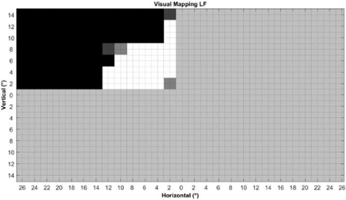
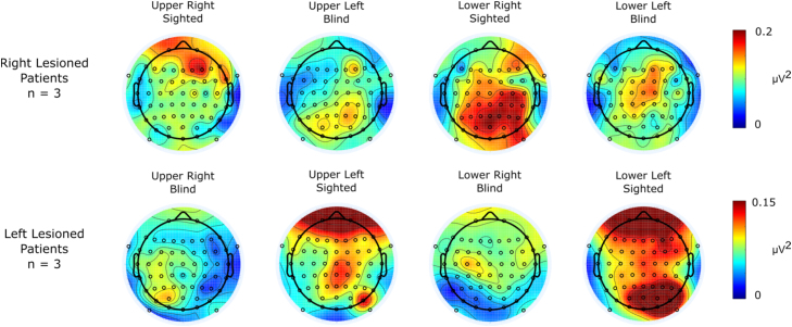
Hemianopia is a visual field defect characterized by decreased vision or blindness in the contralesional visual field of both eyes. The presence of well documented above-chance unconscious behavioural responses to visual stimuli presented to the blind hemifield (blindsight) has stimulated a great deal of research on the neural basis of this important phenomenon. The present study is concerned with electrophysiological responses from the blind field. Since previous studies found that transient Visual Evoked Potentials (VEPs) are not entirely suitable for this purpose here we propose to use Steady-State VEPs (SSVEPs). A positive result would have important implications for the understanding of the neural bases of conscious vision. We carried out a passive SSVEP stimulation with healthy participants and hemianopic patients. Stimuli consisted of four black-and-white sinusoidal Gabor gratings presented one in each visual field quadrant and flickering one at a time at a 12Hz rate. To assess response reliability a Signal-to-Noise Ratio analysis was conducted together with further analyses in time and frequency domains to make comparisons between groups (healthy participants and patients), side of brain lesion (left and right) and visual fields (sighted and blind). The important overall result was that stimulus presentation to the blind hemifield yielded highly reliable responses with time and frequency features broadly similar to those found for cortical extrastriate areas in healthy controls. Moreover, in the intact hemifield of hemianopics and in healthy controls there was evidence of a role of prefrontal structures in perceptual awareness. Finally, the presence of different patterns of brain reorganization depended upon the side of lesion.
偏盲是一种视野缺损,其特征是双眼对侧视野的视力下降或失明。存在大量有记录证明的对呈现给盲视野的视觉刺激的无意识行为反应(盲视),这激发了对这一重要现象的神经基础的大量研究。本研究关注盲视野的电生理反应。由于先前的研究发现瞬态视觉诱发电位(VEPs)不完全适合此目的,因此我们建议使用稳态视觉诱发电位(SSVEP)。阳性结果将对理解意识视觉的神经基础具有重要意义。我们对健康参与者和偏盲患者进行了被动 SSVEP 刺激。刺激由四个黑白正弦光栅组成,一次在每个视野象限中呈现一个,并以 12Hz 的速率逐个闪烁。为了评估响应的可靠性,进行了信噪比分析,并在时间和频率域中进行了进一步的分析,以比较组间(健康参与者和患者)、大脑损伤侧(左和右)和视野(有视力和盲视)。重要的总体结果是,向盲视野呈现刺激会产生高度可靠的反应,其时间和频率特征与健康对照组中皮质外纹状区的特征大致相似。此外,在偏盲患者的完整视野和健康对照组中,有证据表明前额叶结构在知觉意识中起作用。最后,大脑重组的不同模式取决于病变的侧。