Department of Experimental Physics, University of Würzburg, Am Hubland, Würzburg, 97074, Germany.
Comprehensive Heart Failure Center/Deutsches Zentrum für Herzinsuffizienz, University of Würzburg, Würzburg, Germany.
J Cardiovasc Magn Reson. 2017 Oct 16;19(1):77. doi: 10.1186/s12968-017-0382-2.
Local aortic pulse wave velocity (PWV) is a measure for vascular stiffness and has a predictive value for cardiovascular events. Ultra high field CMR scanners allow the quantification of local PWV in mice, however these systems are yet unable to monitor the distribution of local elasticities.
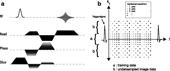
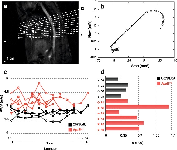
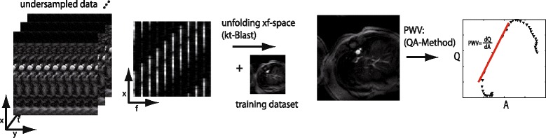
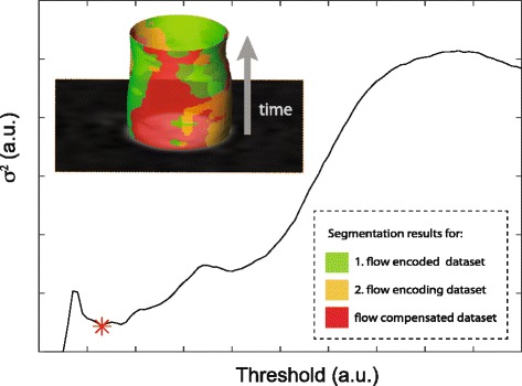
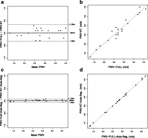
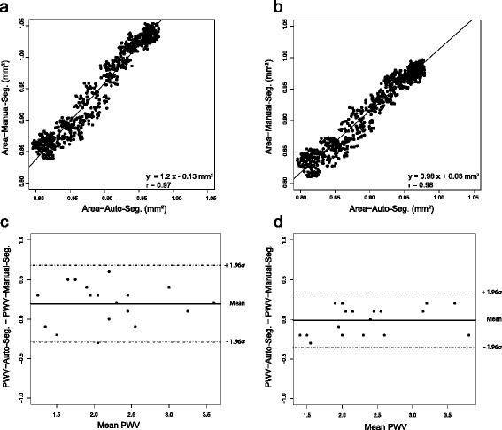
In the present study we provide a new accelerated method to quantify local aortic PWV in mice with phase-contrast cardiovascular magnetic resonance imaging (PC-CMR) at 17.6 T. Based on a k-t BLAST (Broad-use Linear Acquisition Speed-up Technique) undersampling scheme, total measurement time could be reduced by a factor of 6. The fast data acquisition enables to quantify the local PWV at several locations along the aortic blood vessel based on the evaluation of local temporal changes in blood flow and vessel cross sectional area. To speed up post processing and to eliminate operator bias, we introduce a new semi-automatic segmentation algorithm to quantify cross-sectional areas of the aortic vessel. The new methods were applied in 10 eight-month-old mice (4 C57BL/6J-mice and 6 ApoE -mice) at 12 adjacent locations along the abdominal aorta.
Accelerated data acquisition and semi-automatic post-processing delivered reliable measures for the local PWV, similiar to those obtained with full data sampling and manual segmentation. No statistically significant differences of the mean values could be detected for the different measurement approaches. Mean PWV values were elevated for the ApoE -group compared to the C57BL/6J-group (3.5 ± 0.7 m/s vs. 2.2 ± 0.4 m/s, p < 0.01). A more heterogeneous PWV-distribution in the ApoE -animals could be observed compared to the C57BL/6J-mice, representing the local character of lesion development in atherosclerosis.
In the present work, we showed that k-t BLAST PC-MRI enables the measurement of the local PWV distribution in the mouse aorta. The semi-automatic segmentation method based on PC-CMR data allowed rapid determination of local PWV. The findings of this study demonstrate the ability of the proposed methods to non-invasively quantify the spatial variations in local PWV along the aorta of ApoE -mice as a relevant model of atherosclerosis.
局部主动脉脉搏波速度(PWV)是衡量血管僵硬程度的指标,对心血管事件具有预测价值。超高场 CMR 扫描仪可用于量化小鼠的局部 PWV,但这些系统目前还无法监测局部弹性的分布。
本研究提供了一种新的加速方法,可在 17.6T 下使用相位对比心血管磁共振成像(PC-CMR)量化小鼠的局部主动脉 PWV。基于 k-t BLAST(广泛使用的线性加速采集技术)欠采样方案,总测量时间可减少 6 倍。快速数据采集可基于评估血流和血管横截面积的局部时变来量化主动脉血管沿几个位置的局部 PWV。为了加快后处理并消除操作员偏差,我们引入了一种新的半自动分割算法来量化主动脉血管的横截面积。新方法应用于 10 只 8 月龄的小鼠(4 只 C57BL/6J 小鼠和 6 只 ApoE-/-小鼠)的 12 个相邻腹部主动脉位置。
加速的数据采集和半自动的后处理提供了可靠的局部 PWV 测量值,与全数据采样和手动分割获得的结果相似。不同测量方法的平均值没有统计学差异。与 C57BL/6J 组相比,ApoE-/-组的平均 PWV 值升高(3.5±0.7m/s 对 2.2±0.4m/s,p<0.01)。与 C57BL/6J 小鼠相比,ApoE-/-动物的 PWV 分布更为不均匀,反映了动脉粥样硬化病变发展的局部特征。
本研究表明,k-t BLAST PC-MRI 可用于测量小鼠主动脉的局部 PWV 分布。基于 PC-CMR 数据的半自动分割方法允许快速确定局部 PWV。本研究的结果表明,所提出的方法能够非侵入性地量化 ApoE-/-小鼠主动脉的局部 PWV 空间变化,这是动脉粥样硬化的一种相关模型。