Imaging Genetics Center, Stevens Neuroimaging & Informatics Institute, Keck School of Medicine, University of Southern California, Marina del Rey, CA, USA.
Harvard Medical School, Boston, MA, USA.
Mol Psychiatry. 2018 May;23(5):1261-1269. doi: 10.1038/mp.2017.170. Epub 2017 Oct 17.
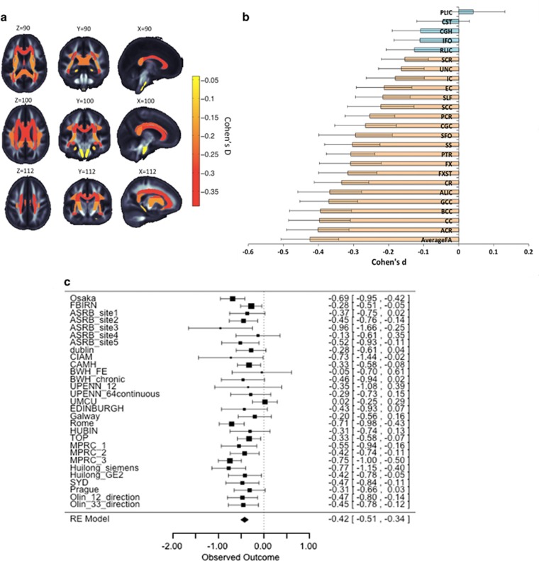
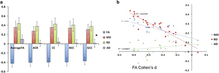
The regional distribution of white matter (WM) abnormalities in schizophrenia remains poorly understood, and reported disease effects on the brain vary widely between studies. In an effort to identify commonalities across studies, we perform what we believe is the first ever large-scale coordinated study of WM microstructural differences in schizophrenia. Our analysis consisted of 2359 healthy controls and 1963 schizophrenia patients from 29 independent international studies; we harmonized the processing and statistical analyses of diffusion tensor imaging (DTI) data across sites and meta-analyzed effects across studies. Significant reductions in fractional anisotropy (FA) in schizophrenia patients were widespread, and detected in 20 of 25 regions of interest within a WM skeleton representing all major WM fasciculi. Effect sizes varied by region, peaking at (d=0.42) for the entire WM skeleton, driven more by peripheral areas as opposed to the core WM where regions of interest were defined. The anterior corona radiata (d=0.40) and corpus callosum (d=0.39), specifically its body (d=0.39) and genu (d=0.37), showed greatest effects. Significant decreases, to lesser degrees, were observed in almost all regions analyzed. Larger effect sizes were observed for FA than diffusivity measures; significantly higher mean and radial diffusivity was observed for schizophrenia patients compared with controls. No significant effects of age at onset of schizophrenia or medication dosage were detected. As the largest coordinated analysis of WM differences in a psychiatric disorder to date, the present study provides a robust profile of widespread WM abnormalities in schizophrenia patients worldwide. Interactive three-dimensional visualization of the results is available at www.enigma-viewer.org.
精神分裂症患者的白质(WM)异常的区域分布仍知之甚少,且不同研究报道的疾病对大脑的影响差异很大。为了在研究中找到共同点,我们首次对精神分裂症患者的 WM 微观结构差异进行了大规模的协调研究。我们的分析包括来自 29 个独立国际研究的 2359 名健康对照者和 1963 名精神分裂症患者;我们协调了各站点间的弥散张量成像(DTI)数据处理和统计分析,并对研究间的效应进行了荟萃分析。精神分裂症患者的各向异性分数(FA)普遍降低,在代表所有主要 WM 束的 WM 骨架的 25 个感兴趣区中的 20 个中都检测到了。效应大小因区域而异,在整个 WM 骨架中达到峰值(d=0.42),主要由外围区域驱动,而不是由定义感兴趣区的核心 WM 驱动。前放射冠(d=0.40)和胼胝体(d=0.39),特别是其体部(d=0.39)和膝部(d=0.37),效应最大。在几乎所有分析的区域中,都观察到了程度较小的显著降低。与扩散测量相比,FA 的效应更大;与对照组相比,精神分裂症患者的平均和径向扩散率显著更高。未检测到精神分裂症发病年龄或药物剂量的显著影响。作为迄今为止对精神障碍 WM 差异进行的最大规模协调分析,本研究为全球精神分裂症患者的广泛 WM 异常提供了一个强有力的特征。结果的交互式三维可视化可在 www.enigma-viewer.org 上查看。